
AI Blood Test Could Detect Early Eye Nerve Damage in People with Type 2 Diabetes
Key Takeaways:
- An AI tool called Pro-DRN uses a blood sample to flag people with type 2 diabetes at high risk of diabetic retinal neurodegeneration (DRN), before any damage shows on the retina.
- It was trained on 1,218 participants and validated in 502 people from UK Biobank, identifying 71 proteins linked to DRN – with ACTA2, COL6A3 and HSPG2 the strongest predictors.
- As retinal nerves are among the first tissues affected by diabetes, the test could also hint at wider nerve damage and help target earlier monitoring and future treatments.
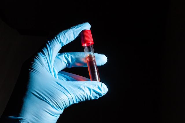
A simple blood test to catch nerve damage early
Scientists have developed an AI-assisted prediction tool that can identify people with type 2 diabetes who are at high risk of developing diabetic retinal neurodegeneration (DRN) before symptoms appear. The findings were published in the journal PLOS Medicine.
The work was led by Wei Wang, MD, PhD, associate professor at the Guangdong Provincial Clinical Research Center for Ocular Diseases. According to the authors, the damage that diabetes inflicts on the delicate nerves of the eye appears to leave a detectable molecular trail in the bloodstream long before it becomes visible in the eye itself.
“Our study suggests that early retinal nerve damage in diabetes leaves measurable signals in the blood,” write the authors. “These findings suggest that a simple blood test analyzed with artificial intelligence may help identify people with diabetes who are at highest risk of early retinal nerve damage, well before visible damage appears on the retina.”
Why the retinal nerves matter in diabetes
Type 2 diabetes affects more than half a billion people worldwide, and it carries an increased risk of long-term complications, including progressive neurodegeneration – the gradual deterioration of nerve tissue over time.
The nerves of the retina are among the earliest tissues to be affected. As this damage advances, it can eventually lead to severe visual impairment and the loss of sight. The difficulty for clinicians is one of timing: current diagnostic methods can only detect DRN once the retina has already sustained irreversible damage. By the time the problem is visible, the window for early, protective intervention has often closed.
How the Pro-DRN tool was built
To address this, Wang and colleagues developed a machine learning algorithm called Pro-DRN. They drew on data from 1,218 participants in the Guangzhou Diabetic Eye Study, all of whom had been diagnosed with type 2 diabetes but had not yet developed DRN at the point of enrolment.
The model combined two distinct streams of information. The first was proteomics data – a detailed read-out of the proteins circulating in participants’ blood samples. The second was a series of yearly retinal images, capturing the state of the eye over a six-year follow-up period. By matching the molecular signals in the blood against how each person’s retina changed year on year, the algorithm learned which blood-borne patterns preceded the onset of nerve damage.
The proteins behind the predictions
The analysis surfaced 71 proteins associated with the development of DRN. Of these, three stood out as the most consistent drivers of accurate prediction: ACTA2, COL6A3 and HSPG2. These are key structural components involved in maintaining the integrity of the nerve and muscle tissue in the eye, which helps explain why disturbances in their levels might signal nerve tissue under strain.
Crucially, the team did not rely on a single dataset. The results were validated in an independent cohort of 502 people from UK Biobank, where the core effects and protein signals were reproduced – an important check that the findings were not simply a quirk of the original group.
From research tool to clinical aid
Pro-DRN has been made available as an interactive, web-based risk assessment tool that clinicians can use to support early DRN screening and to monitor how a person’s risk evolves over time. People identified as being at high risk could then benefit from more frequent check-ups and from early interventions aimed at preventing or slowing progressive neurodegeneration, rather than waiting for damage to become apparent.
A window into the wider nervous system
The potential significance of the test reaches beyond the eye. Because DRN is one of the first signs of nerve degeneration brought on by diabetes, detecting it early could also signal the onset of nerve injury elsewhere in the body.
Such damage can contribute to cognitive impairment, dementia and peripheral neuropathy – the latter causing loss of sensation and motor control in the hands, feet and other extremities. Viewed this way, a single eye-focused test could offer valuable insight into the overall health of a person’s nervous system.
New possibilities for treatment and trials
The discoveries also open up two further avenues. The proteins identified as being involved in DRN progression could be investigated as potential targets for the development of new therapies. In addition, the AI-based tool could prove useful for selecting and stratifying participants in clinical trials that are evaluating neuroprotective strategies designed to prevent or delay nerve damage – helping ensure such studies enrol the people most likely to show a measurable benefit.
Looking ahead
For the researchers, the broader ambition is a shift in how diabetic eye care is approached – from reacting to damage that has already occurred towards anticipating who is most vulnerable.
“Pro-DRN may help move diabetic eye care from detecting established damage toward earlier, molecularly informed risk stratification, so that closer monitoring and future neuroprotective interventions can be directed to the people most likely to benefit,” Wang and colleagues write.
Read More