
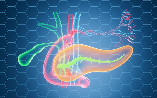
AI Model Detects Diabetes and Sorts Records Into Four Diagnostic Categories
Key Takeaways:
- Researchers have built a machine learning framework that first detects diabetes and then assigns positive records to one of four categories: prediabetes, type 1 diabetes, type 2 diabetes, or diabetes arising from pancreatic disease.
- XGBoost was the authors’ preferred classifier, though the paper reports inconsistent performance rankings across its own tables, with random forest outperforming it in one comparison.
- The framework is a proof-of-concept only. It has not been externally validated, its two stages were trained on separate datasets, and it is not ready for clinical use.
Why diabetes subtyping is a difficult problem
Diabetes is among the most common metabolic conditions worldwide, and its prevalence continues to rise. People living with diabetes typically experience raised blood glucose levels caused by insufficient insulin secretion, insulin resistance, or a combination of the two. Where hyperglycaemia persists, it can lead to serious complications affecting the eyes, heart, kidneys and nerves.
That burden has prompted interest in new strategies for detecting and classifying diabetes using clinical data that is already routinely available. If such tools were externally validated, they could in principle help clinicians identify individuals who warrant further diagnostic assessment, and support decisions about dietary, lifestyle or pharmacological management.
A study accepted for publication in Scientific Reports sets out one such approach: a machine learning (ML) model built on common clinical variables and a derived pancreatic-health index, designed both to detect diabetes and to classify it. The authors are explicit that clinical utility, patient outcomes and quality of life were not assessed.
About the study
The researchers presented an integrated, ML-based approach with two stages. Binary classification was used to determine diabetes status, and multiclass classification was then used to assign records to one of four dataset classes: prediabetes (PD), type 1 diabetes (T1D), type 2 diabetes (T2D), and diabetes from pancreatic disease, also known as pancreatogenic or type 3c diabetes (T3cD).
Two publicly available datasets were used. For the binary task, the team drew on the Pima Indians Diabetes Database, maintained by the National Institute of Diabetes and Digestive and Kidney Diseases (NIDDK), to separate records into diabetic and non-diabetic. For the multiclass task, they used a dataset from the Kaggle repository. The curated multiclass dataset comprised 21,539 samples, split 60% for training and 40% for testing.
How the models were built and tested
Inputs to the multiclass model included age, body mass index (BMI), waist circumference, cholesterol levels, blood glucose levels, insulin levels, and a derived pancreatic-health index.
Several ML algorithms were compared to identify the most effective approach for each classification task: logistic regression, decision trees, random forests, K-nearest neighbours (KNN), naive Bayes, and XGBoost.
To correct class imbalances, the team applied the Synthetic Minority Oversampling Technique (SMOTE) to the training data only. Hyperparameter tuning was then carried out to identify the best-performing parameter combinations, and the models were retrained using those selected parameters. Finally, the researchers ran a Local Interpretable Model-agnostic Explanations (LIME) analysis on their preferred classifier to examine how individual features contributed to model predictions.
What the models achieved
The authors selected XGBoost as their preferred classifier, although the paper’s reported performance rankings are not consistent across tables. Table 2 gives a value of 0.97 for every evaluation parameter, covering accuracy, precision, recall and F1 score. Table 7, however, reports an accuracy of 95.67% for XGBoost against 96.67% for random forest, with random forest also achieving a marginally higher macro-average ROC-AUC, a measure of how well a model discriminates across the four classes.
The researchers attributed XGBoost’s performance to its capacity to capture complex, non-linear associations between features. In the reported test results, XGBoost correctly classified all prediabetes and T1D records, though some confusion persisted between the T2D and T3cD groups. KNN was the least accurate of the algorithms tested.
This kind of discrepancy between a paper’s headline claim and its own supporting tables is exactly the sort of detail clinicians are increasingly expected to spot for themselves. CCH’s CPD-accredited short course AI Essentials for Primary Care covers structured appraisal of AI tools, including the SAFER Evaluation Framework and how to judge AI output against NHS standards.
Which features drove the predictions
General feature-importance analysis pointed to blood glucose levels, insulin and BMI as the most influential variables. The LIME analysis told a slightly different story, indicating that glucose dominated the model’s predictions, with age and cholesterol making secondary contributions in some classes. BMI, waist circumference, insulin and pancreatic health had comparatively lower influence in the LIME results.
Blood glucose levels showed the strongest reported correlation with the class label, at a Pearson’s correlation of 0.86, while insulin showed a correlation of 0.59. These correlations should be treated with caution, since numerical values were assigned to what are, in fact, nominal disease classes.
Age, BMI and waist circumference showed moderate to strong intercorrelations, ranging from 0.63 to 0.68. Their respective correlations with the target were 0.41, 0.46 and 0.56. Pancreatic health showed a negligible negative correlation of -0.06.
In practice, the model primarily learned blood glucose-based decision patterns, which are consistent with the clinical diagnosis of diabetes. The authors interpreted these patterns as broadly concordant with diabetes pathophysiology and existing clinical knowledge, though that interpretation was not independently validated in clinical practice. On the evidence presented, the model is not ready for clinical use and would require considerably more evaluation before it could support, rather than replace, clinical judgement.
Conclusions and future directions
The study demonstrates an ML framework capable of classifying diabetes status and assigning positive cases to four labels: prediabetes, T1D, T2D and T3cD. The authors suggest the framework could eventually assist with diabetes screening and help guide further diagnostic investigation. Crucially, the study did not establish whether using the model improves care, treatment outcomes, quality of life, or the wider global burden of diabetes.
The varying influence of lipid, pancreatic and body measurements may reflect genuine subtype-related biological differences. Equally, it may be an artefact of dataset construction, class coding, correlated predictors, or the absence of clinically verified biomarkers. The authors recommend that these patterns be investigated in clinically characterised datasets before any mechanistic conclusions are drawn.
The framework should currently be regarded as a modular proof-of-concept, because its binary and multiclass stages were trained on separate datasets that may differ in population, variables and collection methods.
Future work, the authors suggest, should use a single training dataset containing both diabetes status and clinically adjudicated subtype labels, including verified pancreatic and autoimmune markers rather than derived variables. External validation in larger and more diverse clinical cohorts would be needed to improve generalisability. Ethical and data privacy concerns would also need to be addressed before any AI-based model of this type could be translated into clinical screening or decision-support settings.
CCH insight
Studies like this one arrive faster than most clinicians can appraise them, and the gap between a promising accuracy figure and a tool that is safe to use in practice is wide. Knowing how to interrogate that gap is now a core professional skill.
AI Essentials for Primary Care is a 100% online, CPD-accredited short course providing 3.5 CPD hours and a Certificate of Completion. It equips the whole primary care team to evaluate AI tools against NHS standards, recognise when AI output should be questioned, and apply the SAFER Evaluation Framework in day-to-day practice. No technical background is required.
Explore AI Essentials for Primary Care →
Read More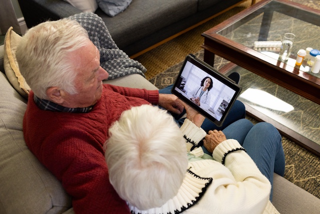
Telemedicine in Nursing Homes Did Not Cut Hospital Admissions, German Trial Finds
Key Takeaways:
- A large cluster-randomised trial across 24 nursing homes in western Germany found no statistically significant reduction in hospital admissions or total days spent in care following an intersectoral, telemedicine-based intervention.
- Researchers tested 1,260 separate model specifications; around two-thirds pointed to a numerical trend towards fewer admissions, but none reached statistical significance (P < .05).
- The authors attribute the null result partly to external shocks, staffing pressures, limited GP engagement and short adaptation windows, and recommend narrower outcome measures and simpler, automated technology in future.
What the study set out to test
A prospective, multicentre cluster-randomised trial has concluded that introducing an intersectoral, telemedicine-based model of care into nursing homes did not produce a statistically significant reduction in hospital admissions among residents.
The research was led by John Grosser, Sophie Pauge, Birthe Aufenberg and Prof. Dr. Wolfgang Greiner of the Department of Health Economics and Health Care Management at Bielefeld University. They worked alongside Miriam Hertwig, Dr. med. Jenny Unterkofler, Dr. med. Christian Hübel and Prof. Dr. med. Jörg Christian Brokmann from the Department for Acute and Emergency Medicine at University Hospital RWTH Aachen, in collaboration with Dr. med. David Brücken of Rhine-Meuse Hospital Würselen and the Optimal@NRW Research Group.
The findings, published in JMIR Aging, evaluate the Optimal@NRW project as it was rolled out across 24 nursing homes in western Germany between May 2021 and April 2023.
The problem the intervention was designed to solve
Optimal@NRW was built around two well-documented pressures in long-term care: non-emergency hospital admissions that could plausibly have been managed in place, and persistent resource shortages across the sector. Transfers to hospital are disruptive for people living in nursing homes, costly for the wider system, and in a meaningful proportion of cases potentially avoidable if clinical assessment and decision support can be brought to the resident rather than the resident to the hospital.
Inside the model of care
The intervention combined four components intended to work as a single, joined-up pathway:
- A 24/7 telemedical consultation centre, giving nursing staff round-the-clock access to remote clinical input.
- Mobile non-physician medical assistants, able to attend the home and carry out assessments in person.
- A virtual emergency hub, coordinating escalation decisions across settings.
- A software-based early warning system for vital signs, designed to flag deterioration before it became acute.
Crucially, the model was intersectoral by design: it was meant to link the nursing home, emergency medicine and primary care rather than sit within any one of them.
A deliberately exhaustive statistical approach
Rather than relying on a single headline model, the research team used specification curve analysis to test how robust any effect was to analytical choices. They evaluated 1,260 distinct mixed-effects regression model specifications, drawing on both primary data collected during the trial and statutory health insurance claims data, with hospitalisation rates and total days spent in care as the outcomes of interest.
The result was consistent across that curve. Roughly two-thirds of the model variations indicated a numerical trend towards reduced hospital admissions, but not one of the 1,260 specifications demonstrated a statistically significant intervention effect at the conventional threshold (P < .05). In other words, the direction of travel was mildly encouraging, but the evidence did not support a claim that the intervention worked.
Why the intervention may not have delivered
The authors are candid that several practical and contextual factors are likely to have compromised the intervention’s chances of success.
External disruptions. The trial period coincided with severe regional flooding in western Germany and with ongoing COVID-19 pandemic restrictions. Both disrupted day-to-day operations in participating facilities and, importantly, distorted baseline hospitalisation rates against which any effect would have been measured.
Staff workload and usability. The model asked a great deal of nursing teams already contending with severe labour shortages. Complex, multi-component tasks – daily manual recording of vital signs among them – added meaningful operational stress rather than relieving it. A digital system that increases the documentation burden on staff is, in practice, competing with the very work it is meant to support.
Physician integration. Engaging general practitioners directly within the telemedical framework proved difficult. Because GP involvement was central to the intersectoral logic of the intervention, that gap limited how far the programme could genuinely connect primary care with the nursing home and the emergency pathway.
Short adaptation windows. Intervention phases ran for between 6 and 15 months per group. The authors suggest this may simply have been too short for staff to embed unfamiliar digital workflows into routine practice, particularly given the competing pressures above.
What the researchers recommend next
The team’s conclusions are constructive rather than dismissive of telemedicine in long-term care. They recommend that future digital health interventions in this setting should:
- Target specific, avoidable admission metrics rather than overall hospitalisations, which are influenced by too many factors outside the intervention’s reach to serve as a sensitive outcome measure.
- Allow extended implementation phases, giving teams realistic time to adapt to new digital workflows.
- Favour simplified, automated solutions such as wearable monitoring devices, reducing the manual data entry burden on nursing staff.
- Pursue deeper structural integration with general practitioners, so that primary care is built into the pathway rather than invited to join it.
What this means for practice
The wider lesson here is one that recurs across digital health evaluation: the technology itself is rarely the binding constraint. Implementation conditions – staffing capacity, workflow design, clinical buy-in and the length of time teams are given to adapt – tend to determine whether a well-conceived tool produces measurable benefit. Practitioners who are asked to assess, adopt or lead on digital tools increasingly need a framework for judging fit and feasibility, not just functionality, which is the ground covered by CPD-accredited training such as CCH’s AI Essentials for Primary Care: Tools, Ethics and Everyday Applications.
For services considering remote monitoring or telemedical support in care homes, the Optimal@NRW findings are a useful corrective. They suggest that ambition should be matched by realism about what frontline teams can absorb, and that outcome measures should be chosen precisely enough to detect an effect if one exists.
CCH insight
Digital tools are arriving in primary and community care faster than most teams can evaluate them. AI Essentials for Primary Care: Tools, Ethics and Everyday Applications is a CPD-accredited short course covering how to appraise digital and AI-enabled tools, work within governance requirements and get reliable results in everyday practice. It carries 3.5 CPD points and counts towards appraisal and revalidation.
Explore AI Essentials for Primary Care →
Source: JMIR Aging
Read More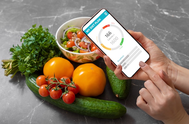
AI Calorie Tracking Apps: Convenient, Popular and Consistently Inaccurate
Key Takeaways:
- Four popular photo-based calorie apps underestimated meal energy by 250 to 345 calories and fat by around 30 grams – roughly a third in both cases.
- Accuracy varied: higher-calorie meals fared better than lower-calorie ones, and carbohydrates were estimated more consistently than other macronutrients.
- Ketogenic meals proved hardest to assess, because their high fat content is consistently underestimated.
Convenience that comes with a margin of error
Advances in artificial intelligence have changed how many people record what they eat. Rather than weighing ingredients or searching a database entry by entry, a person can now photograph a plate of food and let an app estimate its nutritional content in seconds. The convenience is obvious. The accuracy, according to new research, is rather less reliable.
A study conducted at the National Institutes of Health (NIH) Clinical Center compared four popular photo-based apps against meals prepared under tightly controlled laboratory conditions. All four underestimated both calories and fat by approximately a third.
How photo-based calorie tracking works
Photo-based calorie tracking uses AI image recognition to identify the foods present in a photograph of a meal and to estimate the portion sizes on the plate. Those identifications and estimates are then matched against nutrition databases, which the app uses to calculate the energy and macronutrient content of the meal.
The appeal for people managing their weight or monitoring their intake for other health reasons is that the process removes several steps of manual data entry. The trade-off is that the app is making two judgements at once – what the food is, and how much of it there is – before any nutritional calculation begins.
“Photo-based calorie tracking apps are very popular, especially for people trying to manage their health or lose weight,” said Aaron Hengist, a postdoctoral visiting fellow with the Intramural Program of the National Institute of Diabetes and Digestive and Kidney Diseases (NIDDK), part of the National Institutes of Health. “However, the accuracy of many of these apps has not been thoroughly evaluated. Our study helps address this question by looking at whether these apps can reliably estimate calories.”
A metabolic kitchen as the reference standard
The research forms part of a larger diet study at the NIH Clinical Center examining how the body processes nutrients when a person follows either a low-carbohydrate (ketogenic) diet or a standard diet.
Meals for that clinical trial are prepared in a controlled metabolic kitchen – a research kitchen in which every ingredient is weighed to the nearest 0.1 gram. That level of precision gave the investigators something unusual: a set of real, plated meals whose exact nutritional composition was already known.
The researchers took standardised photographs of 102 meals prepared for the diet study and ran the images through four apps: MyFitnessPal, LoseIt!, CalAI and Appediet.
“By using meals prepared in a tightly controlled metabolic kitchen, we were able to compare the apps’ estimates against a precise reference,” said Hengist. “This kind of direct, high-quality comparison hasn’t been available before.”
What the analysis found
Across the 102 meals, all four apps underestimated energy content by approximately 250 to 345 calories on average. Fat was underestimated by around 30 grams. In percentage terms, both calories and fat came in roughly a third below the true values recorded in the metabolic kitchen.
For a person using an app to guide daily intake, an error of this size is not trivial. A consistent shortfall of 250 to 345 calories per meal, repeated across a day, would produce a substantially inaccurate picture of overall energy intake – and would do so in the direction least helpful to someone trying to reduce it.
Where the apps performed better
The findings were not uniformly negative. The analysis showed that MyFitnessPal and LoseIt! estimated the energy content of higher-calorie meals more accurately than lower-calorie meals. All four apps also estimated carbohydrates more consistently than the other macronutrients, suggesting that carbohydrate-containing foods may be easier for image recognition systems to identify and quantify than fats in particular.
Hengist was direct about the practical implication for people using these tools without adjusting the app’s output.
“People using a photo-based tracking app without adjusting the portions or entering the amounts of food should take the results with a grain of salt,” said Hengist. “These apps tend to underestimate calories, especially from fats, so what they actually ate is likely higher than what the app shows.”
Ketogenic meals pose a particular challenge
After completing the initial analysis, the researchers extended the work to more than 200 additional meals in order to understand what factors influence app accuracy.
Their early results suggest that the apps struggle more with meals forming part of a low-carbohydrate ketogenic diet. The likely explanation is straightforward: these meals derive a large proportion of their energy from fat, and fat is precisely the macronutrient the apps most consistently underestimate.
This has implications for anyone supporting people who follow ketogenic or other high-fat dietary patterns, whether for weight management, metabolic health or the management of specific conditions. The tracking tool a person is relying on may be least accurate in exactly the dietary context where fat intake matters most.
For healthcare professionals who regularly review food diaries or app exports during weight management consultations, findings of this kind reinforce the value of structured training in dietary assessment. The College of Contemporary Health’s CPD-accredited Nutrition and Weight Management Essentials short course covers how dietary intake is assessed in practice, the limitations of self-reported data, and how to have constructive conversations with people about what and how much they are eating.
Improving accuracy in the real world
The investigators do not conclude that photo-based tracking should be abandoned. Their view is that combining photo-based features with more traditional methods of measuring diet quality could improve the real-world accuracy of calorie-tracking apps. In practical terms, that means treating the photograph as a starting point rather than a finished record – confirming what the app has identified, adjusting portion sizes, and entering quantities where they are known.
Olivia Charles, a postbaccalaureate intramural research training fellow at NIDDK, will present the findings at NUTRITION 2026, the flagship annual meeting of the American Society for Nutrition, held 25–28 July in National Harbor, Maryland, just outside Washington, D.C. As with research presented at scientific meetings generally, the results should be regarded as preliminary until they appear in a peer-reviewed publication.
For people using these apps day to day, the practical message is a modest one. A photograph is a useful prompt and a low-friction way to build a habit of recording intake. It is not, on the current evidence, a measurement.
CCH insight
Digital tools are increasingly part of how people monitor their diet, but their outputs need to be interpreted rather than accepted at face value – particularly where fat intake or high-fat dietary patterns are involved.
The College of Contemporary Health’s Nutrition and Weight Management Essentials CPD short course equips healthcare professionals with a working understanding of dietary assessment, macronutrient composition and evidence-based weight management, including how to interpret self-reported and app-generated intake data in clinical conversations.
Explore Nutrition and Weight Management Essentials
Source: EurekAlert!
Read More
Stanford Scientists Use AI to Discover a “Natural Ozempic” Free of the Usual Side Effects
Key Takeaways:
- Stanford Medicine researchers used a purpose-built AI algorithm to identify BRP, a 12-amino-acid peptide that reduced food intake in mice and minipigs.
- BRP appears to act specifically in the hypothalamus, and in animal testing did not produce the nausea, constipation or major muscle loss linked to semaglutide.
- The findings remain preclinical, with receptor identification and peptide durability still to be resolved before human trials can begin.
A naturally occurring molecule with semaglutide-like effects
Researchers at Stanford Medicine have identified a naturally occurring molecule that may suppress appetite and reduce body weight in a manner resembling semaglutide, the active ingredient in Ozempic. In animal studies, the molecule also appeared to sidestep several of the problems associated with the drug, including nausea, constipation and substantial muscle loss.
The molecule, known as BRP, works through a different but related metabolic pathway and activates a separate group of neurons in the brain. That distinction could make it a more precise tool for controlling appetite and body weight.
Why a hypothalamus-focused signal matters
The appeal of BRP lies in where it appears to act. Semaglutide’s receptor targets are distributed widely across the body, which helps explain the breadth of its effects – and the breadth of its unwanted ones.
“The receptors targeted by semaglutide are found in the brain but also in the gut, pancreas and other tissues,” said assistant professor of pathology Katrin Svensson, PhD. “That’s why Ozempic has widespread effects including slowing the movement of food through the digestive tract and lowering blood sugar levels. In contrast, BRP appears to act specifically in the hypothalamus, which controls appetite and metabolism.”
The hypothalamus is a small region deep within the brain that helps regulate hunger, body temperature, hormone activity and energy use. Because BRP appears to act mainly in this area, it may influence appetite without producing as many effects elsewhere in the body.
Svensson has co-founded a company that plans to begin clinical trials of the molecule in humans in the near future.
Svensson is the senior author of the research, which was published on 5 March in Nature. Senior research scientist Laetitia Coassolo, PhD, is the lead author of the study.
Hunting for hidden peptides with artificial intelligence
The discovery depended heavily on artificial intelligence, which allowed the researchers to search through proteins belonging to a group known as prohormones.
Prohormones are inactive precursor molecules. They do not perform their final biological function until enzymes cut them into smaller fragments called peptides. Some of these peptides then act as hormones, carrying signals that influence metabolism, appetite and other complex processes in the brain and throughout the body.
A single prohormone can be cut in several different ways, producing many possible peptides. Finding the biologically important ones is difficult, because genuine peptide hormones are relatively rare and can be buried among large numbers of ordinary fragments created during normal protein processing and breakdown.
Traditional laboratory methods can isolate and identify peptides, but the process can generate enormous quantities of data. Researchers may need to sort through hundreds of thousands of molecules to find the few that have meaningful effects.
Narrowing the search to prohormone convertase 1/3
The team concentrated on an enzyme called prohormone convertase 1/3. This enzyme cuts prohormones at specific amino acid sequences and has previously been linked to obesity in humans.
One of the peptides produced through this process is glucagon-like peptide 1, or GLP-1. GLP-1 helps regulate hunger and blood sugar, and semaglutide works by copying its effects in the body.
The researchers reasoned that the same enzyme might produce other peptides that influence energy balance and appetite. To find them, they turned to artificial intelligence.
Peptide Predictor: from 20,000 genes to 373 prohormones
Rather than manually extracting proteins and peptides from tissues and then using methods such as mass spectrometry to identify huge numbers of molecules, the researchers created a computer algorithm called Peptide Predictor.
The program searched all 20,000 human protein-coding genes for the types of sites where prohormone convertases typically cut proteins. The researchers then narrowed the search to genes that produce proteins secreted outside the cell – a common feature of hormones – and that contained at least four possible cleavage sites.
That process reduced the field to 373 prohormones, giving the team a far more manageable group to investigate.
“The algorithm was absolutely key to our findings,” Svensson said.
Peptide Predictor estimated that prohormone convertase 1/3 could produce 2,683 distinct peptides from those 373 proteins. Coassolo and Svensson then focused on the sequences that seemed most likely to affect the brain.
They selected 100 peptides, including GLP-1, and tested whether they could stimulate neuron-like cells grown in the laboratory.
A 12-amino-acid peptide with an outsized effect
As expected, GLP-1 strongly activated the neuronal cells, increasing their activity to three times the level seen in untreated control cells.
One much smaller peptide produced an even more dramatic response. Made from only 12 amino acids, it increased neuronal activity tenfold compared with controls.
The researchers named the peptide BRP after its parent prohormone, BPM/retinoic acid inducible neural specific 2, or BRINP2 (BRINP2-related-peptide).
Amino acids are the basic building blocks of proteins and peptides. A molecule containing only 12 of them is extremely small compared with most full-sized proteins, yet BRP produced the strongest response in the initial cell tests.
Food intake fell by up to 50 per cent
The researchers next tested BRP in lean mice and in minipigs, which more closely mirror human metabolism and eating patterns than mice do.
An intramuscular injection given before feeding reduced food intake during the following hour by as much as 50 per cent in both species.
The team also gave daily BRP injections to mice with obesity for 14 days. On average, the treated animals lost 3 grams, with nearly all of the reduction coming from body fat. Animals in the control group gained about 3 grams over the same period.
The treated animals also showed improved glucose and insulin tolerance. These measures reflect how effectively the body regulates blood sugar and responds to insulin, the hormone that helps move glucose from the bloodstream into cells.
Because incretin-based therapies are now a routine part of weight management for many people living with obesity, clinicians looking to keep pace with the underlying pharmacology and with structured patient support pathways increasingly turn to focused CPD such as the College of Contemporary Health’s GLP-1RA Complete Programme.
No clear signs of the usual side effects
Behavioural testing found no meaningful differences between treated and untreated animals in movement, water consumption, anxiety-like behaviour or faecal output.
The absence of changes in faecal output was especially notable, because semaglutide can slow digestion and cause constipation. The researchers also did not observe the nausea-related responses or major muscle loss associated with some existing weight loss treatments.
Additional measurements of brain activity and body function showed that BRP acts through metabolic and neuronal pathways that differ from those activated by GLP-1 or semaglutide.
Those findings suggest that BRP may reduce appetite through a more focused biological route, although the results remain limited to animals.
What still needs answering before human testing
The researchers are now working to identify the cell-surface receptors that attach to BRP. Receptors are molecular structures that receive signals from hormones, drugs and other chemical messengers. Determining which receptor BRP uses will help scientists understand exactly how the peptide changes appetite and metabolism.
The team also wants to map the full sequence of events that occurs after BRP binds to its target.
Another challenge is duration. Small peptides are often broken down quickly in the body, which can shorten their effects. The researchers are investigating ways to make BRP last longer so that, if it eventually works in people, it could be administered on a more practical schedule.
“The lack of effective drugs to treat obesity in humans has been a problem for decades,” Svensson said. “Nothing we’ve tested before has compared to semaglutide’s ability to decrease appetite and body weight. We are very eager to learn if it is safe and effective in humans.”
CCH insight
Discoveries like BRP sit at the frontier of a field that is already reshaping day-to-day practice. For healthcare professionals supporting people living with obesity, a working command of GLP-1 receptor agonist pharmacology, patient selection, side-effect management and long-term follow-up is now core knowledge rather than a specialism.
The College of Contemporary Health’s GLP-1RA Complete Programme brings this together across three courses and a capstone, leading to an Advanced Certificate, with seven CPD hours and seven downloadable clinical tools designed for use in practice.
Explore the GLP-1RA Complete Programme →
Source: Stanford Medicine
Read More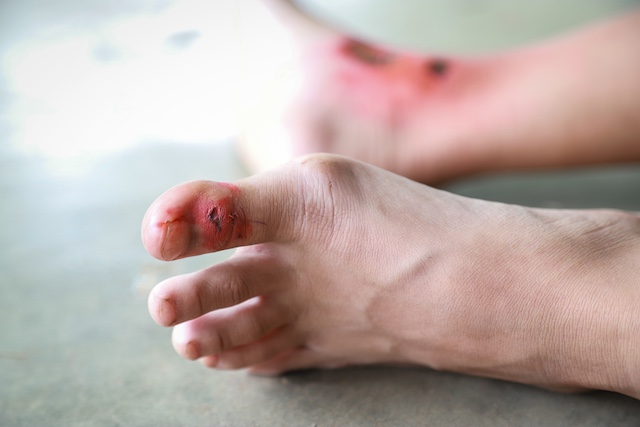
New AI Tool Predicts 12-Week Healing Outcomes for Diabetes-Related Foot Ulcers
Key Takeaways:
- THERMUL is a smartphone-based thermal imaging AI tool designed to predict whether a diabetes-related foot ulcer will heal within 12 weeks.
- The tool was developed and tested in a 110-participant clinical study across two Melbourne hospitals, with images captured at presentation and again at two and four weeks.
- Researchers have secured $490,539 through Australia’s Economic Accelerator and $50,000 from a University of Melbourne fund to validate the technology across metropolitan, regional and remote services.
Turning heat patterns into an early warning signal
A collaboration between the University of Melbourne and RMIT University has produced an artificial intelligence (AI) tool that uses thermal imaging to flag diabetes-related foot ulcers at risk of poor healing, with the aim of reducing hospitalisations and lower limb amputations through earlier intervention.
The tool, named THERMUL, runs on a smartphone and analyses patterns within thermal images of wounds. Those patterns may indicate whether an ulcer is progressing as expected or is at risk of delayed healing. The central output is a prediction of whether a given ulcer is likely to heal within 12 weeks – a window that matters clinically, because ulcers that stall in the early weeks are the ones most likely to progress to serious complications.
Why foot ulceration is such a persistent problem
Diabetes affects over 500 million people globally, and up to 34 per cent of people living with the condition will develop a foot ulcer at some point in their lifetime. Where these wounds are not managed appropriately, the consequences can escalate quickly, running through infection and hospitalisation to lower limb amputation and early death.
The economic picture is equally stark. RMIT University Professor Dinesh Kumar noted that “Diabetes-related foot disease places a significant burden on individuals and the Australian health system, costing an estimated $875 million every year,” positioning the tool as having potential to improve health equity as well as deliver economic benefits for Australia’s healthcare system.
For clinicians working across obesity, type 2 diabetes and cardiometabolic care, complications of this kind sit squarely within the territory covered by the College of Contemporary Health’s CPD-accredited short courses on diabetes and obesity, which examine how metabolic disease drives long-term complications and what earlier, better-coordinated care can achieve.
How the study was carried out
Researchers at the University of Melbourne, RMIT University and Bolton Clarke ran a clinical study at Austin Health and St Vincent’s Hospital Melbourne, involving 110 patients with diabetes-related foot ulcers.
Using a light-weight thermal camera, the team captured images of each ulcer when the person first presented to the clinic, with further images taken two weeks and four weeks later. At the twelve-week mark, clinicians assessed whether each ulcer had healed. That assessment gave the research team a healing outcome to set against the thermal image data collected weeks earlier – effectively a labelled dataset from which an algorithm could learn.
What the algorithm was trained to recognise
Endocrinologist Professor Elif Ekinci, Head of the University of Melbourne’s Department of Medicine and Director of the Australian Centre for Accelerating Diabetes Innovations (ACADI), explained the modelling step directly: “Using this data, we trained an AI algorithm to recognise heat patterns within the initial thermal images that were associated with ulcers that went on to heal, compared with those that did not.”
She was careful to frame the clinical implication as potential rather than proven: “These patterns may provide an early indication of a wound’s likely healing trajectory, potentially allowing clinicians to identify people who might benefit from earlier intervention or escalation of care.”
The distinction matters. Current clinical guidelines often assess healing progress over the first four weeks of treatment to determine whether a diabetes-related foot ulcer is healing as expected. A tool that reads risk from the initial presentation would move that judgement earlier in the pathway, rather than replacing it.
Bringing specialist-level judgement closer to home
One of the clearest arguments for a portable tool is geographic. Regular access to specialist support can be difficult for many people living in regional, rural and remote communities, where the round trip to a specialist diabetes foot service may be measured in hours.
Dr Rajna Ogrin, senior research fellow at Bolton Clarke, set out the gap: “People in regional areas who are living with diabetes and receive care at home, or through outreach services, may not have timely access to specialist diabetes foot services.”
The proposed remedy is a device that travels rather than a person who must: “A portable, non-contact tool like THERMUL has the potential to bring specialist-level decision support closer to where people live and receive care.” The non-contact element is significant in wound care, where minimising physical contact with a compromised wound bed reduces both discomfort and infection risk.
Funding, validation and what comes next
The project initially received seed funding from ACADI, which allowed the research team to take the concept through to prototype stage.
More recently, the team secured $490,539 in grant funding through Australia’s Economic Accelerator programme, alongside $50,000 from the University of Melbourne’s Proof of Concept Fund. That money is earmarked for two purposes: expanding validation of the technology across a wider range of healthcare settings – metropolitan, regional and remote services – and further improving the predictive model using data from more diverse population groups.
THERMUL is currently progressing through the additional validation and development activities required to support future translation into clinical practice. In other words, it is not yet a product in routine use, and the reporting is careful to describe capability in conditional terms.
The partnership behind the tool
Partner organisations include the Australian Centre for Accelerating Diabetes Innovations, Austin Health, Bolton Clarke, St Vincent’s Hospital Melbourne, Royal Flying Doctor Service Victoria and Software Medical Devices Pty Ltd – a mix of academic, acute hospital, community nursing, outreach and commercial partners that reflects the breadth of settings in which diabetes-related foot disease is actually managed.
CCH insight
THERMUL is one of a growing number of AI tools arriving at the point of care, and the practical questions it raises – how a prediction should influence a decision, what validation gaps mean in practice, how to explain an algorithmic result to the person in front of you – are now everyday concerns rather than future ones.
AI Essentials for Primary Care: Tools, Ethics and Everyday Applications is a CPD-accredited short course covering the tools, the ethics and the everyday applications of AI in clinical practice. 3.5 CPD points, 3.5 hours, fully online, start immediately.
Find out more about AI Essentials for Primary Care →
Source: The University of Melbourne
Read More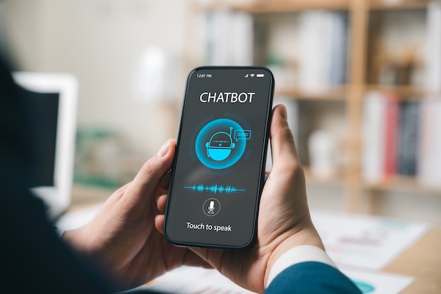
People Like AI Mental Health Chatbots. Whether They Help Is Another Question
Key Takeaways:
- Across 21 studies in 11 countries, people using generative AI mental health chatbots reported high satisfaction and found them convenient and accessible.
- Personalisation and empathy did not reliably translate into better clinical outcomes, and engagement often faded over time.
- The evidence base remains early-stage, leaving safety, equity and crisis response unresolved.
A treatment gap that digital tools are being asked to fill
Generative artificial intelligence (GenAI) chatbots designed to support mental health are winning people over on experience, but the research needed to establish whether they are safe and clinically effective has not kept pace. That is the central finding of a review, currently in press in the journal npj Digital Medicine, which examined the user experience (UX) and intervention design of GenAI mental health chatbots.
The context for this work is a substantial and persistent shortfall in care. Around 25% of people worldwide experience a mental health problem, yet approximately 85% do not receive adequate treatment. The reasons are varied and overlapping: stigma, cost, shortages of trained professionals, geographic distance from services and structural inequities, among others. As the prevalence of mental health conditions has grown while treatment gaps have remained, attention has turned towards innovative models of delivery – and digital tools, with their scalability and convenience, have become a focus of that search.
Digital mental health interventions deliver treatment or support through a range of channels, including chatbots, websites, mobile applications and wearables. Conversational agents, more commonly known as chatbots, are applications that simulate human dialogue using machine learning and natural language processing algorithms.
From scripted responses to open-ended conversation
Traditional mental health chatbots deliver pre-scripted therapeutic content through rules-based or retrieval-based systems. Their strength is predictability, but that same design limits their capacity to personalise support or to recognise what an individual actually needs in the moment.
Chatbots built on large language models (LLMs) work differently. They can simulate core aspects of a therapeutic encounter, including personalised suggestions and empathetic reflections. That flexibility comes with a trade-off. GenAI systems may produce responses that are incorrect or inappropriate, and their open-ended conversational capacity makes intervention design both more consequential and more complex than it is for rules-based systems. When a system can say almost anything, design decisions carry considerably more weight.
How the review was carried out
The researchers set out to map the design characteristics and UX outcomes of interventions involving GenAI mental health chatbots. They began with a systematic literature search to identify studies covering the design and deployment of such tools. Reviews, editorials, media articles and commentaries were excluded.
In total, 21 studies were selected, conducted across 11 countries between 2023 and 2025. The largest numbers came from China and the United Kingdom, followed by the United States. The included work spanned a wide range of maturity, from early-stage prototype evaluations through to clinical trials, with one real-world implementation study.
Most studies recruited general or clinical adult populations, including older people living with dementia. Others involved simulated users or university students. Sample sizes ranged from as few as five participants to as many as 527. Across the body of evidence, there was substantial heterogeneity in outcome measures, and most interventions remained at an early stage of development – two features that shape how much can reasonably be concluded from the literature as it stands.
What the interventions were designed to do
Chatbot interventions most often targeted depression and anxiety, and tended to adopt shared therapeutic mechanisms, including mindfulness, emotion regulation and cognitive restructuring. Some systems were oriented towards mental well-being, stress and loneliness, emphasising general support and preventive care rather than treatment for a specific condition. Others addressed eating disorders, post-traumatic stress disorder and dementia.
The dementia-focused interventions are worth distinguishing. Rather than attempting to address the central neurological features of the condition, they targeted its related psychological dimensions – carer burnout, psychological distress and loneliness among them.
Most interventions were grounded in cognitive-behavioural therapy principles. The specific techniques drawn upon included behavioural activation, psychoeducation, Socratic dialogue, acceptance and commitment therapy, cognitive restructuring and mindfulness.
How the tools were delivered
Interventions varied in frequency, delivery modality and duration. The majority were short-term, running from two to eight weeks. Most were deployed through web-based interfaces and mobile applications, while some were delivered via messaging or social media platforms – meeting people on services they already used rather than asking them to adopt something new.
About 67% of interventions were non-embodied, text-based chatbots. The remainder used voice, avatar-based, augmented reality or other multimodal forms of interaction, with the intention of improving engagement and realism.
What people made of them
All but two of the studies evaluated at least one UX domain. The majority relied on quantitative measures, typically Likert scales, while some gathered qualitative feedback through open-ended questions and semi-structured interviews.
User satisfaction and acceptability were the most commonly reported outcomes. Across studies, participants described the interventions as convenient and accessible, with acceptability generally rated moderate-to-high and reported satisfaction high.
Half of the studies examined usability, using qualitative feedback, the System Usability Scale or Likert scales. Interface design, interaction mode and deployment platform were all observed, alongside differences in usability between studies. A clear preference emerged for free-flowing chat interfaces and customisable features over predefined options. At the same time, some interventions had an unclear scope or limited functionality, leaving people uncertain about what the chatbot could actually do for them.
Usability, engagement and the drop-off problem
Only some studies reported objective utilisation and engagement metrics, such as session frequency, interaction duration, retention over time and task completion. Where these were captured, attrition patterns frequently emerged over time in repeated-measures designs. Uptake in multi-week interventions was often high at the outset before declining – a pattern familiar across digital health more broadly, and one that matters a great deal for interventions whose therapeutic logic depends on sustained practice.
Personalisation and perceived benefit
Most chatbots featured some form of personalisation, reflecting their capacity to adapt conversations and interfaces in response to previous interactions and a person’s emotional state. The most common approach was emotion detection paired with adaptive interaction, allowing people to receive tailored responses and empathetic reflections.
Perceived impact was not consistently measured as a standalone metric. It was more usually folded into qualitative feedback or broader UX evaluations. In the intervention that produced the most granular data, the most frequently reported benefit was improved clarity and awareness.
Where empathy stops being enough
The review’s more cautionary finding is that personalisation and empathy did not consistently translate into stronger clinical outcomes or sustained use. Feeling supported and being helped are not the same thing, and the studies reviewed do not yet demonstrate a reliable link between the two.
Some people reported responses that were repetitive, generic or contextually misaligned. Others raised concerns about over-reliance on chatbots, reduced human contact, data privacy and whether these systems can respond appropriately when someone is in crisis. Inaccurate or clinically misaligned outputs were also linked to an erosion of trust and to disengagement in several studies.
For healthcare professionals, the practical question is less whether these tools have promise than how to appraise them – knowing what a given system is grounded in, where its limits sit and when a conversation needs to move to a human. That judgement is increasingly treated as a core clinical competency, and it sits at the centre of CPD training on the everyday, ethical use of AI in practice.
Design features linked to a better experience
The authors identified several design features associated with better UX outcomes, while being careful to note that these were associations rather than demonstrated causes. They included:
- Deployment on platforms people already knew and used
- Richer interaction modalities beyond plain text
- Integration into existing care pathways
- Personalisation
- Grounding in domain knowledge
- Structured delivery
- Proactive outreach
- Co-design with both experts and end users
The predominance of early-stage studies, combined with limited direct comparative analyses, prevented firm conclusions about which of these features genuinely improved user experience.
What needs to happen next
Taken together, the review suggests that GenAI chatbots have meaningful potential to deliver tailored, empathetic mental health support, and that their acceptability among the people who use them is promising. That is a real finding, and not a small one given the scale of unmet need.
Significant challenges remain, however. Standardising how UX is assessed, grounding intervention design in the needs and preferences of the people who will use these tools, and sustaining engagement beyond the first few weeks all stand out as unresolved. Addressing them, the authors argue, will require co-design with experts and users, validated UX metrics applied in long-term studies, transparent reporting standards, independent evaluation, clearer reporting of model design and training data, and stronger attention to safety, equity and the limits of crisis response.
CCH insight
Generative AI tools are arriving in patient-facing care faster than the evidence base supporting them, which puts the burden of appraisal on clinicians. CCH’s CPD-accredited short course AI Essentials for Primary Care: Tools, Ethics and Everyday Applications covers the practical and ethical judgement this requires – what these tools can and cannot do, where the risks sit, and how to use them safely in day-to-day practice.
Find out more about AI Essentials for Primary Care →
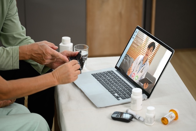
Telehealth Strengthens Diabetes Self-Care but Delivers Only Modest Glycaemic Gains
Key Takeaways:
- Comprehensive telehealth produced modest, non-significant improvements in glycaemic control compared with self-monitoring alone.
- Diabetes self-care was the only outcome to improve significantly, pointing to better self-management rather than better clinical numbers.
- Uptake was low, with relatively few participants completing the intended number of telehealth encounters.
Why self-management sits at the centre of type 2 diabetes care
People living with type 2 diabetes carry much of the day-to-day work of managing their condition. Self-monitoring, medication adherence, lifestyle modification and psychosocial coping all fall largely to the individual, supported at intervals by their clinical team. Comprehensive telehealth has been proposed as a way of closing the gaps between those intervals – enabling more regular contact with health care providers, structured review of patient-generated data, and input from a multidisciplinary team without requiring people to attend in person.
What has remained uncertain is whether that model works in a fee-for-service setting, where reimbursement structures and service design differ markedly from the integrated systems in which much telehealth research has been conducted. A randomised trial published in Annals of Internal Medicine on 23 June 2026 set out to answer that question.
What the trial set out to test
The study, led by Crowley and colleagues, evaluated both the implementation and the effectiveness of a comprehensive telehealth intervention for people with uncontrolled type 2 diabetes and comorbid hypertension, delivered in a fee-for-service context. Participants were randomised either to the comprehensive telehealth programme or to self-monitoring alone, with the comparison designed to isolate the added value of regular provider contact and multidisciplinary review over and above the data-gathering that people were already doing themselves.
Outcomes spanned both the clinical and the behavioural: glycaemic control, blood pressure, weight, diabetes self-care, disease-related distress and self-efficacy, alongside safety monitoring for serious adverse events.
Uptake proved to be the sticking point
One of the clearest findings was not about physiology at all. Uptake of the intervention was limited, with relatively few participants completing the intended number of telehealth encounters. That matters for interpreting everything that follows: a programme that people do not fully engage with is being tested at less than full strength.
Engagement also appeared to shape the results. Improvements in glycaemic control were more evident among participants with greater engagement, suggesting a dose-response relationship that the trial as a whole was not positioned to demonstrate conclusively.
Modest clinical gains that did not reach significance
Compared with self-monitoring alone, comprehensive telehealth was associated with modest improvements in glycaemic control. Those improvements did not reach statistical significance.
The same pattern held across the other clinical measures. The telehealth group showed trends towards better blood pressure and weight outcomes, as well as towards reduced disease-related distress and improved self-efficacy – but again, these differences were not statistically significant. The direction of travel was consistent and favourable; the magnitude simply was not large enough to distinguish the intervention from the comparator.
Self-care was the one clear winner
The exception was diabetes self-care, which was the only outcome that improved significantly with telehealth. It is a result worth dwelling on. It suggests the intervention may strengthen people’s ability to manage their own condition even where clinical measures remain largely unchanged – a benefit that conventional endpoints are poorly designed to capture, and one that may accrue over a longer horizon than the trial allowed.
Whether that improved self-management eventually translates into better glycaemic, blood pressure or weight outcomes is a question the study cannot answer. What it does indicate is that the mechanism telehealth is meant to activate – supported, informed, confident self-management – did in fact activate.
That mechanism depends heavily on the quality of the conversation, not just its frequency. Practitioners looking to strengthen engagement and draw out patients’ own motivation to change are increasingly turning to structured behaviour change training; CCH’s two-hour CPD short course, Behaviour Change Skills: Enhancing Motivation, covers motivational interviewing techniques for exploring readiness, working with ambivalence and recognising change talk in everyday consultations.
Safety and adverse events
Serious adverse events were uncommon and occurred at similar rates in both groups, supporting the safety of the intervention. For a delivery model that reduces face-to-face contact, that reassurance carries weight.
Where the findings stop short
The authors are candid about generalisability. The study population was predominantly low-income with lower educational attainment, and baseline glycaemic control was relatively favourable – leaving less room for improvement than a more poorly controlled cohort would have offered. Digital literacy was not assessed at all, despite being an obvious determinant of who can engage with a telehealth programme and who cannot.
Each of these factors constrains how far the results can be extrapolated to other populations or other service settings.
What this means for practice
The overall picture is one of a promising model that did not, in this setting, demonstrate a clear clinical advantage over self-monitoring alone. Comprehensive telehealth appears capable of enhancing self-management and patient engagement. It did not, here, translate that into measurable clinical benefit.
Further research is needed to establish whether greater uptake of the intervention, or a different patient population, would yield larger benefits. In the meantime, the trial offers a practical reminder for anyone commissioning or delivering remote diabetes care: the technology is only as effective as the engagement it manages to sustain.
CCH insight
This trial found that telehealth improved self-care significantly while clinical measures barely moved – and that engagement was the limiting factor throughout. Motivational interviewing offers an evidence-based way to shift that dynamic, helping people find their own reasons to change rather than pushing against resistance.
Behaviour Change Skills: Enhancing Motivation is a two-hour online CPD course from the College of Contemporary Health, developed with behaviour change specialists at BCT and led by registered dietitians Dympna Pearson and Sam Howard. It carries 2 CPD hours and a Certificate of Completion, and costs £59.
Read More
AI Is Reshaping How Patients Make Healthcare Decisions, Survey Finds
Key Takeaways:
- A majority of US patients now research their symptoms before deciding whether to book an appointment, with AI and search engines central to that process.
- Researchers frame the trend as a global one, driven less by the availability of technology than by mounting frustration with slow, fragmented care.
- The report urges providers to redesign care pathways and deploy their own AI tools to extend capacity and keep patients engaged.
Patients are researching before they reach the clinic
Patients are increasingly taking charge of their own healthcare with artificial intelligence and other digital tools, with 58% of US patients researching their symptoms before deciding whether to book an appointment, according to a new report from the ZS Impact Institute.
The report drew on insights from nearly 10,000 healthcare consumers and providers across the US, Germany and China. Researchers note that the shift is consistent across healthcare systems – a pattern they say points to a “broader breakdown in how patients seek and experience care globally.”
Search engines and AI have become first ports of call
The report found that self-directed research is now routine, and that people who turn to online tools tend to rate them highly. Thirty-seven percent of US respondents report using search engines for health information, 94% of whom find it helpful. Eighteen percent report using AI, and 89% of those describe it as helpful.
The behaviour extends well beyond pre-appointment research. Fifty-two percent of US patients report requesting specific medications, and 68% of providers report seeing an increase in patients requesting therapies by name.
For clinicians, that means fielding more informed – and increasingly AI-shaped – questions in the consulting room. Continuing professional development providers have begun to respond to the trend: the College of Contemporary Health, for example, offers a short CPD course, AI Essentials for GPs, developed to help doctors evaluate AI tools critically and respond with confidence when patients raise them.
Frustration, not just availability, is driving the shift
The report is explicit that the move towards AI “cannot be attributed to availability alone.” Instead, it reflects “growing consumer frustration with healthcare” – a frustration the survey data lays out in some detail.
Forty-five percent of US patients avoid seeing a doctor until they are already unwell, and 41% have not had a health check-up in three or more years. Thirty-six percent wait a year or more for a diagnosis. Once treatment begins, adherence falters: 29% do not take treatments as prescribed, and 54% stop treatments prematurely.
Access is part of the picture too. Thirty-eight percent of respondents report lacking a primary care doctor, and 30% say that diagnostic delays led to a worsening of their medical condition.
What the report recommends
Researchers set out a range of recommendations for leaders across pharma, payers, medtech and providers. For providers in particular, the report suggests redesigning care pathways and using AI to extend both capacity and continuity of care.
Jon Roffman, ZS principal and lead contributor, framed the challenge as one of a system struggling to keep pace with the people it serves.
“Patients are changing faster than the system designed to serve them,” Roffman said in a statement. “AI has put medical knowledge directly in patients’ hands, but the healthcare system still assumes patients will come to it first. That model has changed. Patients are more informed and empowered. To keep them engaged, the system must reduce friction with its own set of AI and technology tools to deliver more connected, supportive care.”
CCH insight
Patients are already walking into consultations with AI-shaped questions and specific therapies in mind. AI Essentials for GPs: Tools, Ethics and Everyday Applications is a three-hour, CPD-accredited short course – created by Prof. Mike Bewick and Dr Dipesh Naik – that gives doctors the frameworks to evaluate AI tools safely, meet NHS governance standards and know exactly what to say when a patient asks. It carries 3.5 CPD hours towards appraisal and revalidation.
Explore AI Essentials for GPs →
Read More
AI Is Now the World’s Top Healthcare IT Investment, Report Finds
Key Takeaways:
- Artificial intelligence is now the leading healthcare IT investment priority across most major markets outside the United States, named by 57% of organisations surveyed.
- Ambient AI dominates clinical interest, with third-party tools such as Microsoft Dragon Copilot commanding the widest cross-regional appeal.
- Cloud adoption is maturing from planning to deployment, with nearly three-quarters of organisations now using the cloud in some capacity.
AI moves to the front of the queue
Interest in artificial intelligence continues to surge, and it has now risen to the top of the priority list for healthcare organisations across most major markets outside the United States. According to a new report from KLAS Research, the share of organisations naming AI as a strategic initiative climbed by 15 percentage points year on year – a striking marker of how quickly the technology has moved from experiment to strategic imperative.
The fourth annual KLAS Research report gathered insights from 182 healthcare organisations spanning 43 countries and territories outside the United States. Artificial intelligence emerged as the single most cited healthcare IT investment priority, named by 57% of respondents. It was followed by electronic health record (EHR) and digitalisation work at 44%, IT and infrastructure at 26%, cybersecurity at 25% and imaging at 23%.
Ambient AI captures the clinical imagination
When respondents were asked which technologies most excited them, ambient AI stood out clearly. A matching 57% pointed to ambient AI as the most “exciting emerging technologies,” ahead of other clinical AI at 17%, operational AI at 16% and general or unspecified AI at 16%.
Governance is keeping pace with enthusiasm. AI strategy, governance and adoption ranked as the leading priority in every region at 37%, followed by clinical documentation at 19%, imaging at 13%, AI platforms and integration at 11%, advanced capabilities at 10%, clinical decision support at 4%, operational efficiency at 3% and patient engagement and experience at 3%.
Among the ambient AI offerings drawing the most attention, the majority are supplied by third-party vendors. Microsoft Dragon Copilot continues to hold the highest cross-regional mindshare, while interest in Heidi Health has grown across Europe and Oceania over the past year. A number of regional players are also in the running, with Augnito attracting interest in the Middle East and TORTUS gaining ground in the UK.
As ambient documentation and clinical AI move from pilot projects into everyday practice, primary care professionals are increasingly expected to understand not only how these tools function, but when their use is appropriate, safe and ethical – a skills gap that structured continuing professional development is beginning to address.
Cloud strategies mature from planning to practice
Cloud adoption is accelerating as organisations shift from planning towards deployment. Nearly three-quarters of respondents now draw on the cloud in some capacity as part of their IT strategy. While only 26% report having moved a core system – such as an EHR, PACS or ERP – into the cloud, organisations worldwide are increasingly operationalising their cloud plans and moving beyond evaluation towards meaningful adoption, according to KLAS data.
Electronic health records sit at the centre of these cloud strategies. More than a third of cloud users – particularly across the Middle East and Canada – have moved all or part of their EHR to the cloud, while nearly a quarter have partially or fully migrated imaging applications such as PACS.
Artificial intelligence is itself shaping how organisations approach the cloud. Those earliest in their cloud journey most often report either using specific AI tools within the cloud or moving their data infrastructure into cloud environments to unlock stronger analytics and computing capabilities.
Electronic health records anchor digital investment
For the organisations reporting EHR and digitalisation investments, nearly a third are either actively implementing an EHR or weighing up replacements or consolidations – chiefly across Europe, the Middle East and Oceania, the report notes.
“Those not involved in such projects are largely focused on increasing digital maturity and optimizing existing EHRs to improve workflows and strengthen the foundations needed for AI and analytics, which is a consistent priority across regions; Latin America in particular has a focus on increasing digital maturity,” the report said.
Consulting demand cools as purchasing slows
Alongside the enthusiasm for AI, analysts observed a cooling in appetite for outside help. Global interest in consulting firms declined between 2025 and 2026, with 42% of organisations reporting no plans to engage such firms and only 25% citing “concrete plans” to bring in consultants for upcoming IT initiatives. The report also noted that 2025 saw the lowest level of EHR purchasing activity in years.
CCH insight
As ambient AI and clinical documentation tools become part of routine care, the professionals using them need to know how to apply AI safely, ethically and effectively at the point of care. The College of Contemporary Health’s CPD-accredited short course, AI Essentials for Primary Care: Tools, Ethics and Everyday Applications (3.5 CPD points, 3.5 hours), is designed to help clinicians do exactly that.
Read More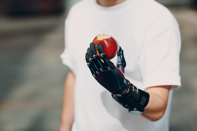
A Smart Exoskeleton Glove Helps People With Hand Paralysis Grasp Everyday Objects
Key Takeaways:
- Researchers in Munich have built a soft, air-powered glove that helps people with hand paralysis grasp everyday objects again.
- Electrical signals from the forearm muscles, read by machine learning, predict a person’s intention to grasp with up to 97% reliability.
- A man living with ALS used the glove to hold a fork for the first time in four years.
A soft exoskeleton driven by air and intention
A soft, pneumatic glove developed at the Technical University of Munich (TUM) is giving people with hand paralysis a way to grasp objects once more. The device was created by researchers at the TUM Chair of Cognitive Systems, who use electrical signals from the forearm muscles to reliably predict the moment a person intends to reach for something. Its designers believe it could one day support people whose hands have been paralysed as a result of accidents or neurological disorders. The research is published in the journal Nature Machine Intelligence.
The team calls the device a “soft-hand exoskeleton”. At its heart is a fabric glove, developed by the researchers, with air cushions fitted to its outer surface. Those cushions are inflated through a total of 13 tubes, each one providing targeted support for the specific hand movements needed to hold a plate or grasp a glass, fork or spoon. Because the cushions can be inflated independently, every finger can be bent and straightened on its own, and the wrist can be rotated too, so that an object can be held securely in the hand.
Reading the intention to grasp
The clever part is knowing when the wearer actually wants to grasp something. To work this out, the researchers measure muscle activity in the forearm. Sensors placed on the forearm capture the faint electrical signals produced by the muscles, and machine learning then analyses those signals to determine the intended movement.
Keeping hold of an object once it has been picked up is a separate challenge. “To prevent objects from being dropped accidentally, we use additional motion sensors to detect transport movements and keep the exoskeleton’s grip securely closed throughout the movement,” says researcher Nicolas Berberich.
Devices like the soft-hand exoskeleton reflect a wider movement of artificial intelligence into everyday clinical care, and professional bodies have increasingly urged healthcare professionals to build the judgement needed to use such tools safely and effectively – the focus of the CPD-accredited digital health training now offered by providers including the College of Contemporary Health.
A soft-hand exoskeleton that anyone can afford
For the team, the appeal of the glove lies as much in its simplicity as in its sophistication. “Our solution is intelligent in two ways,” explains Dr John Nassour. “On the one hand, we’ve developed a highly reliable method of predicting grasping movements by inferring intentions from signals with 97% reliability. On the other hand, with our glove, we’ve developed hardware that optimally supports the intended movements.”
There is a practical advantage on top of that. Dr Nassour sewed the glove himself, and the fabric it requires costs very little. It may not look high-tech at first glance, but it can be used by many people living with paralysis. “We’ve found a solution that anyone can afford but still works very well,” says Prof. Gordon Cheng, director of the Institute for Cognitive Systems.
Central to the project was close collaboration with a man living with amyotrophic lateral sclerosis (ALS).
Working with a person living with ALS
People with ALS gradually lose control of their movements. This happens because the nerve cells responsible for contracting skeletal muscle become damaged and continue to degenerate over time.
By the start of the project, the participant already had very little control over his hands, but he could still move the first joint of his thumb. The researchers built their approach around the strongest signals his thumb muscles could still produce. To record this electromyogram, they attached a sensor to his forearm that picks up the strong signals from the flexor pollicis longus muscle as soon as it moves. Those signals, in turn, trigger the inflation of the glove’s air cushions.
Picking up a fork for the first time in four years
Even though the signals were very weak, the system correctly recognised the participant’s intention in 9 out of 10 cases. With the glove’s support, he was able to reach for objects, hold a fork for the first time in four years, and pick up small cubes and drop them into a container.
A video game played its own part in that progress. The participant had to make a character jump using only the movement of his thumb joint, a simple exercise that helped sharpen the system’s response. The researchers found that just five minutes of this practice was enough to greatly improve his ability to grasp objects. “This patient has shown us that our soft-hand exoskeleton can support him despite one of the most severe neurological disorders,” says Prof. Cheng.
Adapting the glove for more people
The team is now looking beyond ALS. “We are now adapting the concept for other patients, such as stroke survivors,” the researcher adds. A central finding of the current study is that people with severe impairments can more effectively regain the ability to grasp objects with the help of the glove.
That potential is echoed by clinicians working alongside the researchers. Neurologist Prof. Tobias Wächter, from the partner institution Klinik Passauer Wolf, is convinced of what the specialised glove could offer. “In principle, this glove can help people with flaccid paralysis, including, for example, people who have sustained peripheral nerve damage following motorcycle or bicycle accidents, or patients with polyneuropathy,” says Prof. Wächter.
CCH insight
As AI-assisted and digital health tools move from the research lab into everyday practice, the ability to evaluate them safely and confidently is fast becoming a core clinical skill. The College of Contemporary Health’s CPD-accredited short course AI Essentials for Primary Care (3.5 CPD hours) helps nurses, pharmacists, physician associates and the wider team assess new tools, work within clinical governance, and use them responsibly in patient care – no technical background required.
Explore AI Essentials for Primary Care →
Read More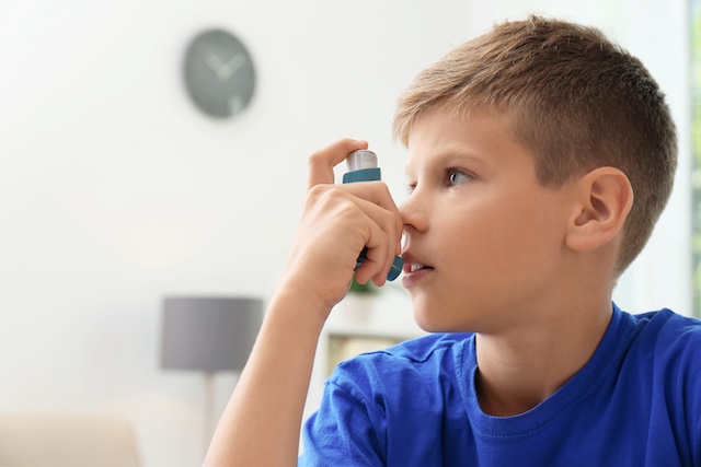
Machine Learning Tool Helps Paediatricians Identify Children at Risk of Persistent Asthma
Key Takeaways:
- A machine learning tool that reads data already held in a child’s electronic health record helped paediatricians more accurately judge which young children are at risk of persistent asthma.
- In a pilot randomised trial using standardised clinical cases, clinicians using the tool reached an average accuracy of 83%, compared with 61% for standard assessment alone.
- The tool is designed to support clinical judgement rather than replace it, and requires no additional tests or questionnaires.
Support for a difficult clinical judgement
A machine learning tool that analyses information already captured in a child’s electronic health record (EHR) has helped paediatricians assess asthma risk more accurately in standardised clinical case scenarios, according to a pilot randomised clinical trial led by a researcher at the Regenstrief Institute. The study was published in the journal Scientific Reports.
The trial evaluated a machine learning-enabled clinical decision support tool known as the Passive Digital Marker. The tool draws on routinely collected EHR data to classify young children as having either a high or a low risk of going on to develop persistent asthma.
Why early asthma risk is hard to predict
Asthma is one of the most common long-term conditions of childhood, yet predicting which young children who have wheezing or other respiratory symptoms will later develop persistent asthma remains difficult. Some children outgrow their early symptoms, while others need ongoing treatment. That uncertainty makes early risk assessment an important, but genuinely challenging, part of paediatric care.
“This tool doesn’t replace a pediatrician’s clinical judgment,” said Arthur H. Owora, PhD, Regenstrief Institute research scientist and lead author of the study. “It helps bring together years of clinical information that’s already in the electronic health record, giving clinicians another source of information when making decisions about a child’s asthma risk.”
How the Passive Digital Marker works
Unlike many prediction tools, the Passive Digital Marker requires no extra testing and asks families to complete no additional questionnaires. Instead, it analyses information that has already been documented in the child’s EHR, including respiratory symptoms, allergies, medication history, respiratory infections and family history. It then presents clinicians with a straightforward high-risk or low-risk assessment.
This approach is intended to save clinicians’ time and reduce the burden on families, since it relies on data that has been gathered over the course of a child’s routine care rather than requiring anything new at the point of decision.
What the trial found
Paediatricians using the tool correctly predicted future asthma more often than those relying on standard assessment alone, achieving an average accuracy of 83% compared with 61%. The improvement was largely driven by better identification of children who went on to develop persistent asthma – the group that is most important to recognise early and hardest to spot.
The researchers stress that the tool is meant to support clinical decision-making, not to supplant it. Its value lies in helping clinicians quickly synthesise years of patient information into a single, easy-to-interpret risk assessment that sits alongside their own expertise. That distinction – between having an AI tool to hand and knowing how to weigh what it tells you – is becoming central to how clinicians are expected to work with these systems.
Limitations and next steps
Because the study used standardised patient cases rather than real-world clinical encounters, further research is needed to establish whether the tool improves outcomes for children in everyday paediatric practice. The pilot demonstrates promise in a controlled setting, but real-world validation is the necessary next stage before wider adoption.
CCH insight
Tools like the Passive Digital Marker are only ever as good as a clinician’s ability to judge when to lean on them and when to look again. That skill – evaluating an AI tool, recognising where it can mislead, and putting sensible governance around its use – is exactly what our short course AI Essentials for GPs: Tools, Ethics and Everyday Applications is designed to build. It’s a 3.5-hour, fully online CPD course led by Prof. Mike Bewick and Dr Dipesh Naik. [Explore the course →]
Funding and authorship
The study was supported in part by the National Institutes of Health under grant K01HL166436. In addition to Owora, it was co-authored by Bowen Jiang, M.S., and Yash Shah, M.S., of the Division of Pediatric Pulmonology, Allergy/Immunology and Sleep Medicine, Department of Pediatrics, Riley Hospital for Children, Indiana University School of Medicine.
Source: Regenstrief Institute
Read More
Physicians May Struggle to Spot AI Errors, Even When Evidence Contradicts the Advice
Key Takeaways:
- In experiments involving decisions about hypothetical patients, physicians tended to trust incorrect advice labelled as artificial intelligence (AI) generated, even when they had the chance to notice that patient recovery data contradicted it.
- Across both experiments, physicians rated the AI system as reliable and did not draw on the recovery data to conclude that its recommendations were wrong; in the second experiment, they failed to notice that the treatment was entirely ineffective.
- The findings, published in the open-access journal PLOS Digital Health by Aranzazu Vinas of the University of the Basque Country and colleagues, point to real challenges for the widely held assumption that a human will reliably catch and correct an algorithm’s mistakes.
What the research examined
New research suggests that physicians may find it difficult to learn from experience when that experience runs counter to advice presented as coming from an AI system. In a series of experiments in which physicians made decisions about treating hypothetical patients, they tended to trust incorrect AI-labelled recommendations, even after being given the opportunity to notice that patient recovery data contradicted those recommendations.
The work was carried out by Aranzazu Vinas of the University of the Basque Country, Spain, together with colleagues, and is published in the open-access journal PLOS Digital Health.
Why AI classification matters in care
AI systems can help physicians categorise patients according to their differing care needs, for example by estimating whether a particular patient is more or less likely to benefit from a given treatment. Because these systems are not perfect, they are intended to be used as suggestions rather than instructions, with any potential errors caught and corrected by the physician using them.
This safeguard rests on an assumption that is easy to take for granted: that a human in the loop will notice when the algorithm is wrong. Prior research, however, has shown that people in general struggle to spot and correct mistakes made by AI. Vinas and colleagues set out to explore how far that difficulty extends to physicians in particular.
How the experiments worked
The researchers analysed data from 223 physicians who took part anonymously in online experiments. Participants were asked to imagine that they had the option to treat patients for a rare disease using a treatment that was not yet proven and still under development. They were told that an AI system had identified which patients were more, or less, likely to benefit from that treatment.
The physicians then chose which patients to treat. After being shown data on how those patients recovered, they rated their perceptions of how reliable the AI system was.
The design contained a deliberate mismatch. The actual effectiveness of the hypothetical treatment did not align with the AI’s recommendations. In the first experiment, the treatment was equally, and moderately, effective for all patients. In the second experiment, it was equally ineffective for everyone. In each case, the recovery data available to physicians should, in principle, have allowed them to see that the AI’s classification did not hold up.
What the physicians did
In both experiments, the physicians tended to rate the AI system as reliable, and they did not appear to use the patient recovery data to conclude that the AI’s recommendations were incorrect. In the second experiment, they did not realise that the treatment was entirely ineffective.
As lead author Aranzazu Vinas notes: “In both experiments, physicians mostly trusted the AI’s classifications and had trouble learning from the feedback. Furthermore, in the second experiment, professionals did not notice that the treatment was completely ineffective.”
Co-author Helena Matute adds: “People tend to say that there is always a human controlling the algorithm, but our experiments show that doctors (as well as anyone else) have problems in learning from the available evidence when it contradicts the suggestions of an algorithm.”
What it means for healthcare
Taken together, the results highlight potential challenges for incorporating AI-based classification into healthcare. If the human overseeing an algorithm cannot readily detect its errors, even when contradicting evidence is in front of them, then the reassurance that a clinician will always catch a mistake may be weaker than commonly assumed.
The authors suggest that future research could build on this study, for instance by developing and testing strategies and protocols designed to strengthen human critical thinking and the detection of AI errors. The aim would be to maximise the benefits of human-AI collaboration while minimising the potential for error.
Co-author Fernando Blanco summarises the wider purpose of this line of enquiry: “It is important to investigate the errors that humans (including doctors) make when working with algorithms, in order to learn how to minimize the problems that arise from them.”
Building the habit of questioning AI
While researchers work on formal protocols, individual clinicians can already sharpen how they interrogate AI output. Knowing when to trust a recommendation, and when to challenge it, is a clinical skill rather than a technical one, and it is one that structured training can help build. Our short course AI Essentials for GPs: Tools, Ethics and Everyday Applications introduces practical frameworks, including the SAFER Evaluation Framework, for spotting errors, fabrications, and outdated recommendations before they reach a patient. For clinicians who want a reliable method for the kind of critical checking this study suggests is all too easy to skip, it is a useful place to start.
Read More