

Child sleep apnea severity linked to obesity, finds new study
Researchers at UT Southwestern Medical Center and Children’s Health have discovered that obesity and older age are substantial predictors of the severity of obstructive sleep apnea (OSA) in children. The study, recently published in Laryngoscope Investigative Otolaryngology, underscores the essential role of obesity in paediatric OSA. This new finding contributes to the increasing recognition of the association between childhood obesity and various negative outcomes including cognitive deficits, impaired academic performance, behavioural issues, excessive daytime sleepiness, and potential long-term effects on cardiovascular health.
Obstructive sleep apnea is a sleep disorder characterised by repeated obstruction of the upper airway during sleep, leading to interruptions in both ventilation and sleep patterns. Diagnosis of OSA usually requires full-night polysomnography, an extensive sleep study that monitors brain waves, blood oxygen levels, and heart and breathing rates while the patient sleeps.
Traditionally, paediatric patients suffering from OSA have been treated with adenotonsillectomy, a surgical procedure involving the removal of the adenoids and tonsils. However, this surgery does not always resolve the problem, and some children continue to experience what is known as residual OSA after the procedure.
The study was led by Romaine F. Johnson, M.D., M.P.H., Professor of Otolaryngology – Head and Neck Surgery at UT Southwestern, who emphasised that “OSA is typically considered a condition that affects adults.” But Dr. Johnson pointed out that the growing epidemic of childhood obesity has led to an increase in the number of paediatric patients diagnosed with OSA. He explained that “higher levels of obesity and advancing age are key contributors to this issue,” and suggested that targeting childhood obesity could substantially enhance sleep quality and mitigate the detrimental consequences of both OSA and residual OSA.
Dr. Johnson and his research team initially embarked on the study to investigate the relationship between socioeconomic status (SES) and paediatric OSA. They examined the medical records of 249 children aged 18 or younger who underwent both polysomnogram and adenotonsillectomy at Children’s Health over a one-year span. To evaluate the influence of SES on paediatric OSA, the researchers utilised the area deprivation index (ADI), a metric defining neighbourhood-level disadvantages through various social determinants such as education, employment, and poverty.
Contrary to expectations, the study revealed that neighbourhood-level deprivation, as indicated by the ADI ranking, did not predict OSA severity or residual OSA after surgery. Instead, the team found that severe OSA was significantly associated with obesity, and residual OSA was more commonly linked with older age, particularly among adolescents.
While the research did not establish a direct link between SES and OSA severity, Dr. Johnson emphasised the need for further investigation into the relationship. He highlighted the critical nature of understanding how socioeconomic status intersects with paediatric OSA, stating that “social disadvantages can further affect the consequences of poor sleep quality.” He also noted that children who experience both OSA and social disadvantages might be at increased risk and may require specialised interventions.
The significance of this study lies in its identification of childhood obesity as a key factor in the severity of sleep apnea, thereby opening the door to potential interventions targeting weight management. Supported by the Beth and Marvin C. “Cub” Culbertson Professorship in Pediatric Otolaryngology, the research also emphasises the complex interplay between obesity, age, and sleep disorders in children, and calls attention to the need for a multifaceted approach to address these interconnected health challenges.
Read More
Luton Council takes action against childhood obesity by banning unhealthy food ads on municipal property
Luton Council has made a significant decision to combat the growing concern of childhood obesity in the town by implementing a ban on advertising for unhealthy food items on most of its council-owned property. As part of this determined initiative, advertisements featuring food products high in fat, salt, and sugar will no longer be permitted to be displayed on billboards, lampposts, screens, or roundabouts within the town’s jurisdiction.
This move, according to the council, aims to “protect children and adults” from the influence of marketing that promotes unhealthy eating habits. The situation has become particularly alarming in Luton, where childhood obesity rates have exceeded national levels. The latest figures from the National Child Measurement Programme (NCMP) revealed that 11.4% of reception age children in Luton (ages four to five) were classified as having obesity, compared to the national rate of 10.1%. Similarly, obesity among children aged 10 to 11 (Year 6) in Luton stood at 29.2%, noticeably higher than the national rate of 23.4%.
Labour councillor Khtija Malik, who holds the portfolio for public health, declared the ban as a “start” in the town’s efforts to decrease its “high rates of obesity among young children.” Ms. Malik stressed the importance of fostering healthy eating habits early in life, acknowledging the considerable impact advertising has on shaping people’s food choices.
This ban, although extensive, will not encompass all advertising locations within the town. Unhealthy food adverts may continue to be seen on bus shelters until at least 2027, as the council’s current advertising contract for these spaces extends to that year. However, Ms. Malik reassured that the council, having “control of our assets,” would exercise control over “what is advertised” in areas under its jurisdiction.
The move by Luton Council has not only demonstrated a proactive approach to a pressing public health issue but has also been met with support from advocacy groups. Fran Bernhardt, the children’s food campaign coordinator at Sustain, a group dedicated to better food and farming, lauded the council’s efforts, stating, “Luton Council has stood up to the food and drinks industry on behalf of all their residents.” She further emphasised the importance of the decision, pointing out that those residing in the most deprived areas are the ones most affected by unhealthy food advertising and are consequently most at risk from diet-related diseases.
The decision by Luton Council signifies an important step in recognising and addressing the complex factors that contribute to childhood obesity. By limiting the exposure to advertising that encourages unhealthy food choices, the council is actively working to create an environment that supports healthier lifestyle decisions. This ban is a notable example of local governance taking decisive action to influence public behaviour and health, reflecting a growing awareness of the pivotal role advertising plays in shaping dietary habits and preferences. It serves as a landmark measure that could potentially inspire similar initiatives in other towns and cities, contributing to a broader societal shift towards prioritising public health.
Read More
Hypothalamic differences discovered in people who are living with overweight or obesity
A groundbreaking study by researchers at the University of Cambridge has provided new insights into the role of the hypothalamus in controlling appetite. This small yet critical region of the brain, which is about the size of an almond, appears to have structural differences in those who are living with overweight or obesity compared to those of healthy weight.
Dr. Stephanie Brown from the Department of Psychiatry and Lucy Cavendish College, University of Cambridge, noted the importance of the hypothalamus in regulating appetite but also acknowledged the difficulty in studying it in living humans due to its small size.
The study team addressed this challenge by using a machine learning algorithm to analyse MRI scans of 1,351 young adults across different BMI categories. Published in Neuroimage: Clinical, their findings revealed that the hypothalamus’s overall volume was significantly larger in those who with overweight or obesity.
These findings offer further evidence to the connection between brain structure, weight, and food consumption, as obesity is a global concern affecting over 1.9 billion people worldwide. The condition increases risks for numerous health problems, including type 2 diabetes, heart disease, stroke, and mental health issues.
The team also explored the possibility that the changes in the hypothalamus might be related to inflammation. Previous animal studies have shown that a high-fat diet could cause inflammation in the hypothalamus, leading to insulin resistance and obesity. In some cases, just three days of a fat-rich diet was enough to trigger inflammation.
Dr. Brown emphasised that if similar processes occur in humans, a high-fat diet could impact our ability to sense when we’ve eaten enough, leading to weight gain. The researchers hypothesised that the brain might react to inflammation by enlarging specialised immune cells, known as glia, causing the hypothalamus to be larger.
Professor Paul Fletcher, the study’s senior author, expressed hope that this new approach to analysing brain scans would deepen our understanding of appetite control in obesity. While the exact implications of these findings are still unclear, more research is needed to confirm whether the increase in hypothalamic volume is a cause or result of being overweight, or if those with larger hypothalami are naturally inclined to eat more.
Supported by the Bernard Wolfe Health Neuroscience Fund, Wellcome, NIHR Cambridge Biomedical Research Centre, and additional funding from Alzheimer’s Research UK, the study presents a transformative view of obesity. It opens new avenues for exploring obesity from a neurological perspective, potentially revolutionising how obesity and overweight conditions are understood and treated. Future studies may provide essential insights into intervention strategies, reshaping the way we approach this global health challenge.
Read More
Obesity linked to impairment of immune cells protecting against psoriasis
Obesity is more than just a condition associated with excess weight; it can also influence the immune system in ways that make the body more susceptible to certain diseases. Recent research has shed light on how obesity may impair immune cells that protect against inflammation associated with psoriasis, a common skin disorder. This newfound understanding could pave the way for new treatments for psoriasis, particularly for those who suffer from obesity.
Psoriasis, marked by an accumulation of dry, scaly skin patches, is found to be twice as prevalent in individuals with obesity as compared to those without. This connection between obesity and psoriasis has long been observed, but the underlying mechanisms have remained elusive.
Researchers at Emory University in Atlanta, Georgia, led by Chaoran Li, took up the challenge to explore the intersection between obesity, inflammation, and psoriasis. They specifically investigated regulatory T-cells, which play a crucial role in controlling inflammation. These cells maintain a delicate balance in the skin, offsetting pro-inflammatory immune cells. While obesity is known to increase inflammatory cells, its influence on regulatory T-cells was not well understood.
Li’s team conducted a detailed genetic analysis of regulatory T-cells in various organs of mice, including the skin, spleen, lungs, and lymph nodes. They identified a unique subset of these cells that were prevalent in the skin of the rodents. Interestingly, this group was also found to be abundant in human skin samples.
The research took a significant turn when scientists genetically modified mice to lack these specific cells. When they applied a cream that induces psoriasis-like symptoms, such as skin thickening, the modified mice’s skin became nearly 50% thicker in a week compared to those with the anti-inflammatory cells. This observation strongly suggested that these particular cells have a role in preventing psoriasis symptoms.
To delve into the relationship with obesity, another group of mice were fed a high-fat diet for 16 weeks. The skin of these mice with obesity displayed fewer regulatory T-cells and was more reactive to the psoriasis-inducing cream than the skin samples from mice on regular diets. This correlation between obesity and a decrease in anti-inflammatory immune cells that prevent psoriasis was a pivotal discovery.
These findings not only highlight obesity’s impairment of crucial immune cells but also hint that a high-fat diet may be a key factor in psoriasis and other skin-related inflammations. This insight opens up the possibility that dietary interventions could be a viable treatment option for psoriasis.
Another promising avenue explored by the researchers involved the drug pioglitazone, known to boost the activity of regulatory T-cells. It was found to reduce skin inflammation in mice with obesity. Intriguingly, pioglitazone is also a type 2 diabetes medication. Some studies have indicated that individuals with diabetes on this drug have a lower risk of psoriasis, raising the potential of the medication as a dual-purpose treatment.
However, the applicability of these findings to humans still requires validation. Li’s team plans to examine how this specific subset of cells varies in humans with both psoriasis and obesity compared to those with one or neither condition.
This research represents a significant stride in understanding the intricate relationship between obesity, immune function, and psoriasis. By unearthing the cellular mechanisms behind this connection, it could potentially lead to more targeted and effective treatment options for those struggling with psoriasis, especially in the context of obesity. It also emphasises the importance of a healthy diet and its impact on overall skin health.
Read More
Rising obesity prevalence may have altered perceptions of healthy body weights, study suggests
A concerning trend has emerged, revealing that an increasing number of overweight teenagers do not perceive themselves as being too heavy. This phenomenon raises alarms as it may lead to detrimental lifestyle choices, warn scientists. The trend of underestimating body weight was found to be particularly prevalent among girls, according to a peer-reviewed study.
The study suggests that the rising prevalence of obesity and the emergence of body trends idealising an athletic, muscular physique may contribute to teenagers underestimating their weight and becoming resistant to adopting healthy diet and exercise habits. However, the researchers acknowledge that other factors, including body image, dieting, changing eating patterns, and migration, may also play a role in these shifting perceptions over time.
Conducted by the International Health Behaviour in School-Aged Children, the study surveyed 11 to 15-year-olds between 2002 and 2018, analysing trends in body weight perception while considering age, gender, and family socioeconomic status. The survey encompassed over 745,000 adolescents from 41 countries across Europe and the United States. These findings are particularly significant for the UK, where more than a third of teenagers have either overweight or obesity.
Lead author of the study, Dr. Anouk Geraets from the Department of Social Sciences at the University of Luxembourg, emphasised the potential impact of body weight perception during this impressionable age on teenagers’ lifestyle choices, including their food consumption and exercise habits. Dr. Geraets expressed concern regarding the trend of fewer adolescents perceiving themselves as overweight, as this could undermine efforts to address the escalating obesity levels among this age group. Teenagers who underestimate their weight may not recognize the need to lose excess weight and may consequently make unhealthy lifestyle choices.
The study revealed an increase in underestimation of weight status and a decrease in overestimation over time, with girls being more susceptible to these perceptions than boys. Scientists worry that shifting trends in perception could diminish the effectiveness of public health interventions targeting young people.
Dr. Geraets emphasised the clinical and public health implications of the study. While the increase in accurate weight perception and the decrease in overestimation may have a positive impact by reducing unnecessary and unhealthy weight loss behaviours among adolescents, the rise in underestimation highlights the need for interventions to strengthen accurate weight perception. Further research is necessary to understand the underlying factors driving these trends and to develop effective public health interventions.
The study’s findings were published in the journal Child and Adolescent Obesity, shedding light on the urgent need to address teenagers’ misperceptions of their weight in order to promote healthier lifestyles and combat the growing obesity crisis among this age group.

Study challenges belief that moderate alcohol consumption shields against obesity and type 2 diabetes
Moderate alcohol consumption does not provide protection against conditions such as obesity and type 2 diabetes, according to a recent study, challenging the long-debated notion of potential health benefits associated with light to moderate drinking.
The study investigated whether modest alcohol intake had any positive effects on health—a subject that has divided experts. Researchers analysed data on alcohol consumption from over 400,000 participants and discovered a higher risk of obesity and type 2 diabetes among individuals who consumed more than 14 drinks per week. Notably, this link was more pronounced in women than in men.
Dr. Tianyuan Lu of McGill University in Québec commented on the findings, stating, “Previous research has suggested that moderate drinkers may be less susceptible to developing obesity or diabetes compared to non-drinkers and heavy drinkers. However, our study indicates that even light-to-moderate alcohol consumption, defined as no more than one standard drink per day, does not safeguard against obesity and type 2 diabetes in the general population.”
The study’s results revealed that heavy drinking was associated with increased measures of obesity, such as body mass index, waist-to-hip ratio, and fat mass, as well as an elevated risk of type 2 diabetes.
Importantly, the study did not find any evidence of improved health outcomes related to moderate alcohol consumption in individuals who consumed seven or fewer alcoholic drinks per week.
Dr. Lu concluded by highlighting the significance of the research in promoting awareness of the risks associated with alcohol consumption. The study’s findings aim to inform future guidelines and recommendations regarding alcohol use, with the hope of encouraging healthier behaviours as alternatives to drinking.
For further details, refer to the full study published in JCEM.
Read More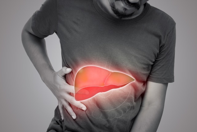
UK’s soaring liver cancer death rate blamed on alcohol and obesity
Liver cancer deaths in the UK have seen a staggering 40% increase over the past decade, making it the fastest-growing cause of cancer-related fatalities in the country, warns the British Liver Trust. Calling attention to the alarming rise, the charity is urging immediate measures to address the “carcinogenic effects” of inexpensive alcohol and unhealthy food.
Pamela Healy, CEO of the British Liver Trust, identifies alcohol and obesity as the key drivers behind the surge in liver cancer cases and deaths. She stresses the need for government intervention to combat these issues, asserting that such action can create a healthier environment for all. Healy emphasises the urgency of addressing the accessibility and affordability of unhealthy food, often priced significantly lower than nutritious alternatives.
This call for action comes amidst delays in implementing regulations that would prohibit supermarket multi-buy deals on high-fat, high-sugar, or high-salt foods. The Association of Directors of Public Health has also urged ministers to introduce minimum unit pricing on alcohol, following Scotland’s successful implementation of a minimum price of 50p per unit in 2018.
The British Liver Trust has submitted evidence to the parliamentary health and social care committee’s inquiry into future cancer care, focusing on innovative approaches to diagnosis and treatment. In addition to advocating for earlier detection and improved access to effective therapies, the charity emphasises the importance of prevention. Liver disease, a significant risk factor for liver cancer, can be reversed through lifestyle modifications, including reduced alcohol consumption and weight loss.
The submission by the British Liver Trust emphasises the need for upstream interventions to mitigate the carcinogenic impact of an unhealthy food and beverage environment. The charity cites evidence indicating that population-wide measures regulating the affordability and accessibility of alcohol and unhealthy food are more effective in reducing disease burden and addressing health inequalities than individual behaviour change alone.
The charity calls for a comprehensive alcohol strategy that tackles promotions and affordability. It highlights the fact that alcohol is classified as a group 1 carcinogen, known to cause at least seven types of cancer. Data from Cancer Research reveals that liver cancer mortality rates per 100,000 people in the UK have risen from 6.4 in 2007-2009 to 9.3 in 2017-2019. Primary liver cancer has one of the lowest survival rates among all cancers.
The British Liver Trust emphasises the need for enhanced surveillance of high-risk groups with liver disease to enable early diagnosis and improve survival rates. Additionally, the charity advocates for increased investment in liver cancer research.
The UK government is facing mounting pressure to take more effective action to combat obesity. Danone UK and Ireland, one of the country’s major food companies, recently suggested that the government should consider taxing products high in fat, sugar, or salt to tackle the obesity crisis.
In response to the inquiry, the Department of Health and Social Care affirms its commitment to diagnosing 75% of cancers at stages 1 and 2 by 2028. The department outlines its interventions, including community health liver checks for those at high risk of cirrhosis, with a particular focus on deprived communities.
A spokesperson from the Department of Health and Social Care highlights ongoing efforts to encourage healthier food and drink choices and tackle obesity. Recognising that obesity costs the NHS approximately £6.5 billion per year and is the second leading cause of cancer, the spokesperson highlights initiatives such as calorie labelling in restaurants, cafes, and takeaways to empower individuals to make informed lifestyle choices.
The spokesperson further notes that the NHS has witnessed record numbers of cancer patients in recent years, with earlier-stage diagnoses becoming more prevalent. To support this, the government is strengthening the cancer workforce and has published the first-ever Long-Term Workforce Plan, backed by over £2.4 billion, aiming to deliver the most extensive training expansion in NHS history over the next 15 years.
Read More
New study links obesity to 18 types of cancer, including leukaemia and lymphoma
In a concerning development, a recent study has established a direct connection between excess body weight and an elevated risk of developing multiple types of cancer. While previous research had identified 13 cancers associated with overweight or obesity, this groundbreaking study expands the list to include 18 different forms of the disease. The findings highlight the urgent need for a reevaluation of the impact of overweight and obesity on cancer rates and emphasise the significance of weight management from an early age.
The study, conducted by researchers in Catalonia, Spain, examined the electronic health records of over 2.6 million individuals aged 40 or older who were initially cancer-free. Surprisingly, within a span of just nine years, more than 225,000 participants received a cancer diagnosis. The researchers meticulously analysed the data, paying close attention to individuals with a body mass index (BMI) of 25 or greater, indicative of having overweight or obesity.
Published in Nature Communications, the study’s results reveal a troubling correlation between a BMI of 25 or higher and an increased susceptibility to 18 distinct types of cancer. What is particularly noteworthy is that the risk of cancer development begins as early as 18 years old. Furthermore, individuals who were overweight during their early adulthood (between the ages of 18 and 40) faced a significantly higher risk of developing cancer. The risk further intensified for those who struggled with excess weight for extended periods and those who experienced severe obesity in their youth.
Dr. Heinz Freisling, a co-author of the study, emphasised the implications of these findings, urging a reassessment of the current estimation of the cancer burden associated with overweight and obesity. It is highly likely that the actual burden is underestimated due to these newly discovered connections.
This study adds to the growing body of evidence highlighting the long-term health risks stemming from childhood obesity, including its impact on cognitive function and brain structure. Additionally, the researchers identified several types of cancer that are now linked to excess weight, including leukaemia, non-Hodgkin lymphoma, head and neck cancers, and bladder cancer.
Previously recognised connections between excess weight and cancer include adenocarcinoma of the oesophagus, postmenopausal breast cancer, colon and rectal cancer, uterine cancer, gallbladder cancer, gastric cardia cancer, kidney cancer, liver cancer, ovarian cancer, pancreatic cancer, thyroid cancer, meningioma (a type of brain cancer), and multiple myeloma, as reported by the Centers for Disease Control and Prevention (CDC).
Dr. Panagiota Mitrou, director of research, policy, and innovation at the World Cancer Research Fund, emphasised the public health implications of this comprehensive study. The new findings establish connections between overweight/obesity and additional cancers, such as leukaemia and non-Hodgkin lymphoma. Maintaining a healthy weight throughout life, starting from early adulthood, emerges as one of the most critical preventive measures against cancer.
The study underscores the need for proactive public health measures, focusing on early prevention and weight management strategies to reduce the incidence of these cancers and promote overall well-being. By prioritising healthy weight maintenance, individuals can significantly mitigate their risk of developing various types of cancer and safeguard their long-term health.
Read More
Exercise and weight loss combination amplifies battle against obesity and prediabetes
Groundbreaking research conducted at Washington University School of Medicine in St. Louis reveals the tremendous impact of combining regular exercise with a 10% body weight loss in combating obesity and prediabetes. The study found that this combination more than doubles insulin sensitivity compared to weight loss alone, reducing the risk of developing Type 2 diabetes and coronary heart disease.
The researchers initially knew that modest weight loss improves insulin sensitivity, and exercise alone has limited efficacy in aiding individuals with obesity and prediabetes in shedding pounds. However, they were astonished to discover the profound effect that combining a consistent exercise regimen with a 10% reduction in body weight can have on insulin sensitivity.
Published in the journal Nature Metabolism, the findings shed light on the crucial role of exercise in improving overall health outcomes for individuals struggling with obesity and prediabetes.
Dr. Samuel Klein, the senior investigator and director of the Center for Human Nutrition, explained the significance of the study’s results: “Insulin resistance is a major factor that causes Type 2 diabetes, nonalcoholic fatty liver disease, and abnormal blood lipids in people with obesity. We’ve shown that combining exercise with weight loss causes a marked improvement in whole-body insulin sensitivity, thereby lowering the risk of developing diabetes and treating obesity-related metabolic diseases to a much greater degree than is possible with weight loss alone.”
The study involved 16 participants with a body mass index (BMI) ranging from 30 to 49, indicating obesity. All participants had prediabetes and demonstrated medical evidence of insulin resistance.
Eight participants underwent a diet-only intervention, resulting in a 10% body weight loss. The other eight individuals followed the same dietary plan but also engaged in supervised exercise sessions multiple times per week.
Dr. Klein highlighted the significance of their findings: “The data from most studies show that exercise has very little effect on body weight in people with obesity… Our study involved detailed analyses of metabolic changes in muscle and body fat before and after a 10% weight loss in people who lost weight with diet therapy alone and in those who lost the same amount of weight with diet therapy plus supervised exercise training. The results demonstrate that the benefits of combining exercise with weight loss are considerable.”
With over 37 million Americans living with diabetes and more than 40% of the population grappling with obesity, which is a significant contributor to diabetes cases, the study’s findings have far-reaching implications. Approximately 96 million adults in the United States, or one in three, are estimated to have prediabetes.
Dr. Klein emphasised the crucial role of exercise in weight management therapy, stating, “The metabolic benefits we found in this study demonstrate the profound reasons why exercise should always be included in weight-management therapy.”
Read More
Revitalising weight loss: The miraculous power of “beige fat” in reversing age-related weight gain
Discovering the ability to activate the body’s own fat-burning mechanisms and counteract age-related metabolic changes might seem like an unattainable dream, but researchers at Cornell University’s Division of Nutritional Sciences are working towards making it a reality. Their recent study explores the potential of a specific type of fat cells known as “beige fat” to reverse the effects of a slowing metabolism.
In mammals, including humans, two main types of fat exist: white adipose tissue (WAT), which stores excess calories, and brown adipose tissue (BAT), which burns calories to generate heat and regulate body temperature. The study sheds light on the therapeutic possibilities of a third type of fat called beige fat, a subtype of WAT. Beige fat shares characteristics with both white and brown fat, possessing thermogenic properties similar to brown fat and the ability to regulate blood sugar levels and reduce fatty acids associated with conditions like arterial hardening and heart disease.
The formation of beige fat occurs when adipose progenitor cells, a type of stem cell found within white fat, are stimulated by prolonged exposure to cold temperatures. However, as individuals age, their response to this stimulus weakens, leading to an imbalance favouring the production of white fat.
“Seasonal changes in beige fat occur in young humans, but an older person would have to stand outside in the snow in their underwear to achieve those same effects,” explains Dan Berry, an assistant professor in the Division of Nutritional Sciences at Cornell University.
Previous research by Berry revealed that the ageing process inhibits the formation of beige fat cells in response to cold temperatures.
The study’s lead author, Abigail Benvie, a doctoral student researcher in Berry’s lab, explains that the ultimate goal of their research is to find ways to stimulate metabolic pathways without subjecting individuals to prolonged cold exposure. Researchers successfully suppressed this pathway in ageing mice and stimulated the production of beige fat cells in areas where only white fat would typically form.
This research not only offers potential solutions for age-related weight gain and associated health conditions but also provides insights into the molecular mechanisms underlying beige fat formation. The study’s co-authors, including graduate students Derek Lee, Benjamin M. Steiner, and Siwen Xue, as well as Yuwei Jiang from the University of Illinois at Chicago, plan to further investigate the identified pathway and explore other molecular regulators of beige fat formation. With a $2.2 million, five-year grant from the National Institutes of Health, Berry’s lab aims to deepen our understanding of how these regulators change in levels and activity during the ageing process.
The study, published in the journal Nature Communications, represents a significant step forward in harnessing the potential of beige fat to combat age-related weight gain and improve metabolic health.
Read More
Family-focused approach in paediatric primary care proves effective for child weight loss
A groundbreaking family-based treatment for childhood obesity, developed by researchers at the University at Buffalo (UB), has demonstrated significant success in a multicenter study conducted in paediatric primary care settings. Previously available only in specialised clinics, this evidence-based treatment has now been implemented for the first time in four U.S. cities, targeting children aged 6-12 and their parents.
Published in the Journal of the American Medical Association, the study reveals that family-based treatment conducted in paediatricians’ offices leads to improved weight-loss outcomes not only for the treated child and parent but also for untreated siblings. The research, supported by the National Heart, Lung and Blood Institute, emphasises that the healthy behaviours promoted during the treatment extend beyond the immediate family members, potentially improving the overall health of the entire family.
In the study, three times as many children in the treatment group (27%) experienced a clinically meaningful reduction in median body mass index (BMI) compared to the usual care group (9%). These improvements in weight correlated with enhanced cardiometabolic outcomes, such as blood pressure, lipids, and glucose regulation. The success of this novel family-focused treatment, pioneered by senior author Leonard H. Epstein, has significant implications for the long-term health and well-being of the treated children.
The family-based treatment program involves the implementation of eating and activity plans, education in parenting and behavioural techniques, and the facilitation of support for positive behaviour changes within both the family and peer environments. In individualised sessions, parents and children are seen together, reviewing self-monitored eating and activity levels, treatment manuals, handouts, and setting goals for weight and behaviour.
The randomised clinical trial enrolled 452 children aged 6 to 12, with one parent, in primary care practices across Buffalo, Rochester, Columbus, and St. Louis. Half of the children received family-based treatment, while the other half received usual care. Notably, the treatment demonstrated positive outcomes across racial and ethnic backgrounds.
Children receiving family-based treatment showed a significant difference in percent over median BMI compared to those receiving usual care, with a reduction of 6.48%. Furthermore, parents experienced a reduction of 3.97% in BMI, and untreated siblings who were overweight had a reduction of 5.38%. These changes in BMI among family members were interrelated, suggesting that the treatment fosters the modelling of healthy behaviours and facilitates real change in the shared family environment.
The success of this behavioural intervention underscores the growing need for similar treatments in paediatric primary care. The study authors emphasise the necessity for “coaches” in primary care settings who can provide support for obesity treatment and address various behavioural issues, including anxiety, depression, and eating disorders.
The implementation of this family-focused approach not only ensures the immediate health of children but also equips them with lifelong healthy practices. The study’s positive outcomes highlight the potential for transforming paediatric primary care into a hub for comprehensive behavioural interventions, addressing a wide range of health-related concerns.
Read More
Study reveals obesity as a precursor to mental disorders across all ages
A recent study conducted by the Medical University of Vienna in collaboration with the Complexity Science Hub Vienna has uncovered that individuals suffering from obesity face an elevated risk of developing various mental disorders across all age groups. Interestingly, the study reveals that women are more susceptible than men to most of these disorders. The research findings were published in Translational Psychiatry, a renowned scientific journal.
The researchers meticulously analysed a comprehensive dataset, encompassing all inpatient hospitalisations in Austria between 1997 and 2014. The aim was to ascertain the relative risks of coexisting diseases in individuals with obesity and to pinpoint any statistically significant variations between genders. The data illustrated that an obesity diagnosis markedly heightens the likelihood of developing an array of mental disorders, including depression, nicotine addiction, psychosis, anxiety, eating disorders, and personality disorders.
Michael Leutner, who headed the study and is part of the Department of Internal Medicine II at MedUni Vienna, asserted, “The findings highlight the imperative need for increased vigilance toward psychiatric diagnoses in patients with obesity and engaging specialists early on if necessary.”
Elma Dervic, the co-first author of the study from the Complexity Science Hub, expounded on the innovative approach adopted to discern which illness typically manifested before or after an obesity diagnosis. “This novel method enabled us to identify trends and common patterns in the onset of diseases,” Dervic explained. The study revealed that, with the exception of disorders in the psychosis spectrum, obesity was likely to be diagnosed before the emergence of any psychiatric condition.
Contrary to a widely-held belief among physicians that psychopharmacological medications were responsible for the connection between mental disorders and obesity, the study suggests otherwise. “While this might hold true for schizophrenia, our data does not corroborate this for depression or other mental disorders,” elucidated Alexander Kautzky, the first author from the Department of Psychiatry and Psychotherapy at MedUni Vienna. However, it remains uncertain whether obesity directly impacts mental health or if early stages of psychiatric disorders are frequently overlooked.
Notably, the study found women to be more predisposed to nearly all mental disorders, except for schizophrenia and nicotine addiction. Approximately 16.66% of men with obesity were found to have nicotine addiction compared to just 8.58% of women with obesity. Conversely, the rate of diagnosed depression was nearly threefold higher in women with obesity (13.3% in women with obesity compared to 4.8% in women who did not have obesity) than in men with obesity (6.61% in men with obesity compared to 3.21% in men who did not have obesity).
With over 670 million individuals affected by obesity globally, it is well-established that obesity contributes to metabolic disorders and severe cardio-metabolic complications such as diabetes mellitus, arterial hypertension, and dyslipidemia. The revelation that obesity is frequently a precursor to serious mental disorders underscores its significance as a health risk factor, particularly for younger populations where the risk is most pronounced.
Consequently, the researchers stress the urgency for comprehensive mental health screening amongst patients with obesity to enable timely preventive measures or the provision of appropriate treatment.
Read More